Oral Flora
I-INTRODUCTION:
For many decades, the oral cavity was simply considered the entrance to the digestive tract. Then, based on the wealth of new data establishing a bidirectional and reciprocal relationship between general health and the ubiquitous bacteria of the Oropharyngeal sphere, the concept of the Oral Ecosystem was introduced.
The oral environment is a physicochemical environment, which occupies and influences the oral cavity as a compartment.
This oral environment therefore includes the anatomical structures which limit it (buccal mucous membranes, tongue, teeth), salivary secretions (and by extension the glands which produce them), the oral immune system, and the flora which colonizes this space.
II-GENERAL INFORMATION:
1-DEFINITION OF BACTERIA:
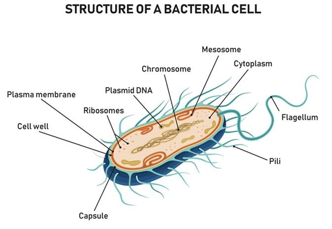
Bacteria are single-celled prokaryotic organisms measuring 1 to 10 µm, comprising:
-Constant elements: nucleus, cytoplasmic wall and cell membrane.
-Inconstant elements: spores, cilia, flagella, glycocalyx and capsule.
2-COMPOSITION:
1-The bacterial wall: is a rigid structure which protects the bacteria from the high osmotic pressure within the cytoplasm and has antigenic power.
– The Gram + bacteria wall: is composed of 90% peptidoglycan (murein) with many carbohydrate bonds. There are also acids ( lipoteichoic acids (LTA) which are connected to the lipids of the plasma membrane and which give solidity to the cell.
– The Gram-positive bacteria wall:
Peptidoglycan represents less than 20% of the weight of the bacteria. The wall is divided into two structures: the outer membrane and the periplasmic space.
- The outer membrane is a phospholipid bilayer (which contains LPS: Lipopolysaccharide Acid). It has: porins (transport of hydrophilic molecules (such as antibiotics), proteins (transport of growth factors and iron), and structural proteins.
LPS: All bacterial endotoxins are lipopolysaccharides according to Rietschel and Brade 1992), are composed of two parts: an antigenic part (surface antigen: the specific polysaccharide chain) of the molecule; and a lipid part anchored in the external membrane, is responsible for toxicity and activation of the host immune response by macrophages.
-Only free LPS molecules not anchored in the bacterial cell wall are toxic to the host, they are generated (by the formation of vesicles, or during cell division, or during the dissolution of the cell wall of dead bacteria).
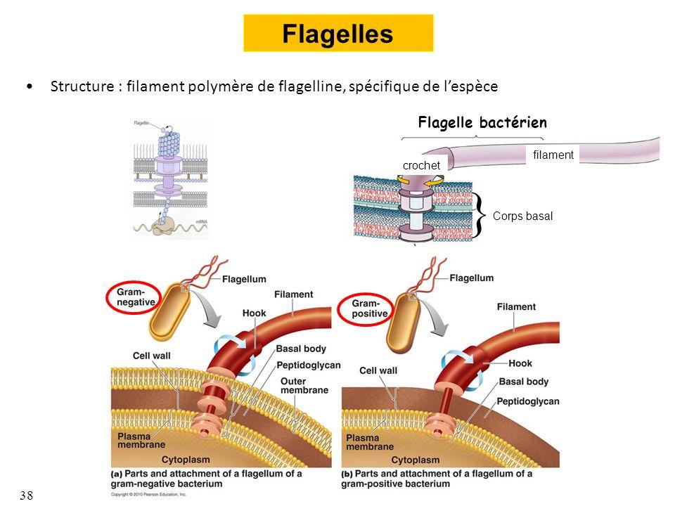
- The plasma membrane: is composed of proteins surrounded by a phospholipid bilayer. It plays a role in the exchange of metabolites thanks to permeases and the production of ATP. It also serves as an attachment to flagella.
- Flagella: They play a role in mobility, chemotaxis and antigenicity, allowing the identification of the bacteria.
- Fimbriae: Are short filamentous structures found on the surface of Gram-positive and Gram-negative bacteria. Are made up of the protein fimbrillin.
In addition, they are thin structures whose main function is to attach the bacteria to its substrate. They therefore play a role in the adhesion of bacteria to the host’s epithelial cells and in the virulence of certain bacteria.
Pili: we distinguish two types:
Sex pili: Longer and in smaller numbers (rarely more than three), found on the surface of Gram-negative bacteria, made up of pilin proteins. They play an essential role in bacterial conjugation (responsible for horizontal gene transfer).
Common pili (fimbriae): are filamentous protein structures. They play a role in attaching bacteria to surfaces.
- Capsule: The capsule is a polysaccharide structure surrounding the wall of Gram-negative bacteria. It prevents phagocytosis of bacteria, and promotes adhesion.
- Glycocalyx : This is a polysaccharide envelope on the surface of the bacteria that promotes its adhesion to host cells and makes it resistant to antiseptics and antibiotics.
- Spores: Some bacteria, such as Bacilli, can form endospores when the environment becomes poor in nutrients. These are inactive, non-pathogenic forms of the bacteria that are highly resistant to temperature changes and chemical agents, such as antiseptics or antibiotics. However, when the environment becomes favorable again, these spores give rise to identical bacteria.
Note: Fimbriae, glycocalyx and lipoteichoic acid: are bacterial mediators of adhesion.
3-CLASSIFICATION:
3-1-according to the Form:
- Cocci (spherical) which can be in a chain or in a cluster
- Bacillus (elongated rod shape)
- Coccobacillus (oval)
- Filamentous
- Spiral
- Vibrio (curved)
2-according to Gram staining:
This coloring was developed by Hans Christian Gram in 1884 (use of two dyes (gentian blue and fuchsin : pink)
– so-called gram+ bacteria: turn blue-violet
– so-called gram bacteria: turn pink
It allows us to distinguish between the two types of bacterial walls, which have a great influence on the infectious power and sensitivity of bacteria to antibiotics.
Gram-positive bacteria are resistant to the decolorization of gentian violet by alcohol and therefore appear purple under the microscope.
On the other hand, Gram-negative bacteria, due to the high lipid content of their wall, allow alcohol to pass through and are therefore discolored. They appear pale pink after re-staining with fuchsin .
3-Breathing mode :
- Strict aerobic: can only live in the presence of oxygen
- Micro-aerophile: optimal growth in an environment where the oxygen concentration is lower than the atmospheric concentration.
- Capnophile: optimal growth in a medium enriched with CO2 .
- Optional: can use different breathing modes
- Strictly anaerobic: can only live in the absence of oxygen.
III-DEFINITION OF ORAL FLORA (ORAL MICROBIOTA):
The oral microbiota is the set of microorganisms present in the oral cavity. These microorganisms are essentially bacteria, about 700 species. Viruses that are transient (herpes), yeasts (candida albicans), protozoa and archaea are also present. Long called oral flora.
This so-called saprophytic flora is distributed according to the different surfaces to be colonized: saliva, the tongue and the different mucous membranes, the subgingival site and the dental surface.
The oral microbiota is acquired at birth from maternal and environmental microbiotas. These microbiotas colonize the child’s oral cavity. The eruption of temporary teeth drastically modifies the existing ecosystem. New surfaces with different physicochemical characteristics must be colonized. Overall, bacterial diversity and richness increase and diverge from the maternal microbiota during the development of the oral cavity and from the age of 2 years would be close to those observed in adults.
IV-COMPOSITION OF THE ORAL FLORA:
-THE SUPRA GINGIVAL FLORA:
It is dominated by facultative anaerobic germs, while spirochetes are rare (0.1%), streptococci dominate, also neisseria and veillonella.
-THE SUBGINGIVAL (SULCULAR) FLORA:
There is an increase in the number of strict anaerobes: Bacteroides, Fusobacterium, Peptostreptococcus, Porphyromonas prevotella and Aggregatibacter.
-THE FLORA OF THE LANGUAGE:
The tongue is an important niche for microbial germs, it is dominated by facultative anaerobic streptococci, we find there: streptococcus salivarius (dominant), veillonella, actinomyces, bacteroides, peptostreptococcus .
V-BACTERIAL ADHESION:
Adhesion is defined by the ability of bacteria to attach themselves to a surface. It is the set of phenomena that oppose the separation of two bodies in contact. Without this property, no bacteria could multiply or cause pathological phenomena (Mouton and Robert, 1994)
Adhesion can be summarized in four successive dynamic stages:
1-TRANSPORT:
Bacteria approach PAE through three mechanisms:
Passive diffusion : this is the movement that drives all bacteria and allows them to move randomly .
Motility: it is defined by the bacteria’s own movements, thanks to the presence of flagella on its surface. This mechanism can be associated with chemotaxis, which allows bacteria to approach surfaces .
Convection: it results from the movements of the tongue and salivary flows .
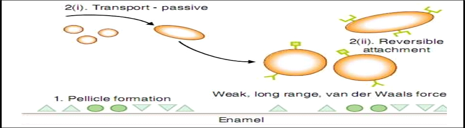
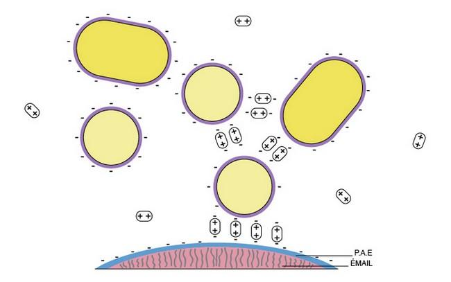
2-NON-SPECIFIC INITIAL MEMBERSHIP:
The bacteria and the surface begin to interact with each other when the distance between them is 50nm through physicochemical interactions:
– REPULSIVE FORCES:
Enamel proteins and acquired pellicle proteins are rich in polar acid groups (negative charge), the bacterial surface contains acid groups (negative charge), which generate electrostatic repulsive forces.
-ATTRACTIVE ELECTRODYNAMIC FORCES OF VAN DER WAALS:
Bacteria are subject to attractive forces whose core of attraction is greater than that of the repulsive forces.
The repulsion is counterbalanced by the electrodynamic attraction (VANDER WALLS force) so the resultant has the effect of maintaining the bacteria at a certain distance from the substrate (reversible phase)
– FORMATION OF CALCIUM BRIDGE: (irreversible phase):
The approach of the bacteria to the dental surface is favored by the establishment of a calcium bridge between the negatively charged surfaces (tooth-bacteria).
– HYDROPHOBIC INTERACTIONS:
They come into play during adhesion in an aqueous medium. Water molecules are arranged in an ordered structure, assimilated to apolar molecules, thus being able to form bonds with other apolar molecules. These interactions established between molecular sequences of a hydrophobic nature present on the surface of the bacteria and the substrate ensure a stable environment that allows other weak interactions to occur: we then speak of positive cooperation.
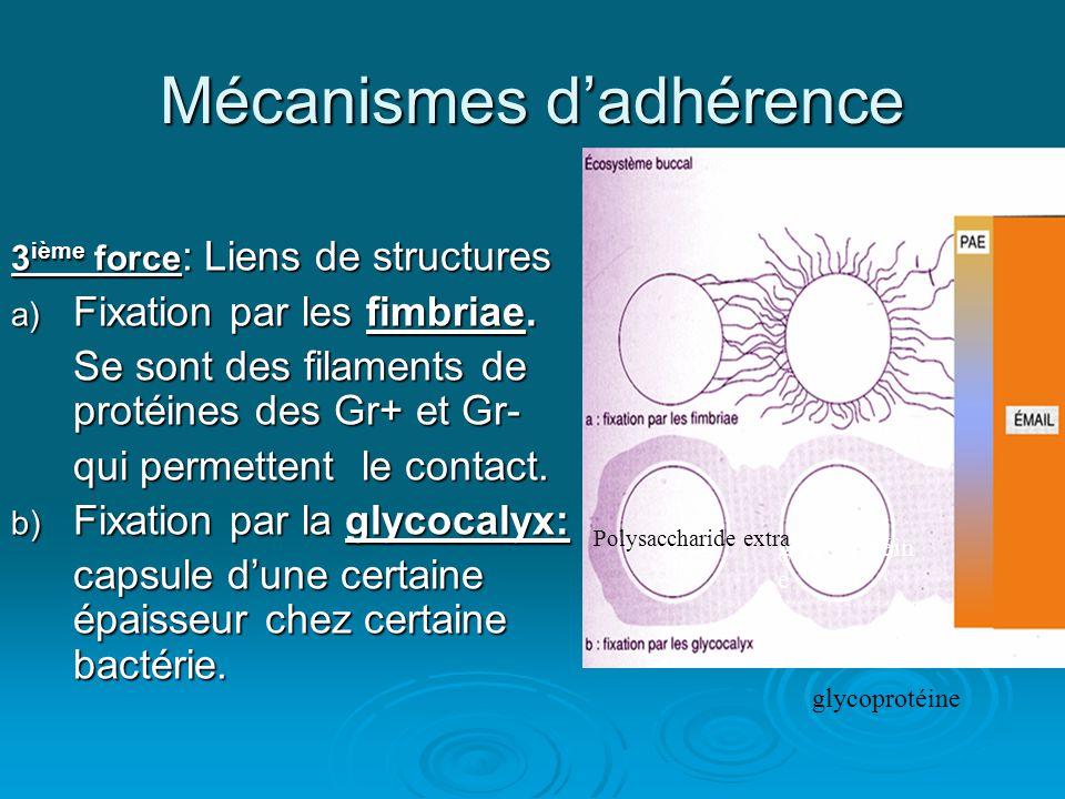
-FIXATION BY GLYCOCALYX:
The glycocalyx is composed of polysaccharides or glycoproteins and secreted by the bacteria and surrounds it, giving it a hydrophilic character. The glycocalyx can fill the space between bacteria and substrate, and the adhesins it contains allow irreversible fixation.
-FIXATION BY THE FIMBIAE:
They are extracellular appendages, made up of polymerized proteins in the form of filaments, the filament carries adhesins on the lateral glycoprotein chains.
By establishing a bridge between the bacterial body and the surface to be colonized, the fimbriae allow contact to be established, although the bacteria are still at a distance from their attachment substrate; and as soon as contact has taken place, the fimbrial adhesins keep the bacteria close to the surface, allowing new forces of attraction to come into play, such as hydrogen bonds.
3-ATTACHMENT:
To remain on dental surfaces for a long period of time, bacteria form high-affinity bonds, using specific surface molecules:
(SPECIFIC INTERACTIONS):
-Adhesin-Receptor type interactions: generally ensured by the adhesins contained in the fimbriae and the glycocalyx in the ligand-receptor form, in fact each bacterial adhesin molecule specifically recognizes its receptor on the surface to be colonized (dental or mucous membrane).
-Enzyme – Substrate type interactions:
For example, S. mutans produces enzymatic complexes: Glycosyltransferases (GTFs). These allow the formation of glycans in the presence of sucrose. These glycans, which are very sticky, bind specifically to GTFs, present on the surfaces of other streptococci, or to receptors present on PEA. Among the PAE receptors, at the level of dental surfaces, we find mainly proteins rich in histidine, proline (PRP), lysozyme and α-amylase.

4-COLONIZATION:
When the microorganisms are firmly attached, growth can begin and the bacteria can multiply.
Pioneer bacteria are able to synthesize many surface exopolysaccharides to allow new species to co-adhere and form micro-colonies on dental surfaces. The PEA receptors are then all saturated and colonization enters a phase of slow bacterial multiplication.
VI-TRANSFORMATION OF SAPROPHYTIC FLORA INTO PATHOGENIC FLORA:
Under the influence of several factors: (increase in the number of germs, arrival of exogenous germs, increase in the virulence of germs, weakening of the host’s defenses), this microbial flora will cause lesions in the oral cavity such as dental caries and periodontal diseases; but a selection of germs will take place to result in one of the diseases.
Factors influencing the growth of microorganisms:
*Physicochemical factors: temperature T°, PH, presence of O2 and humidity
*Host factors : diet, host defense, hormonal change (pregnancy)
*Genetic factors: appear to influence intestinal and oral flora
*Age: from 70 years old: increase in staphylococci (S. aureus), lactobacilli, actinomyces naeslundi.
And from the age of 80: the increase in the number of yeasts (candida albicans)
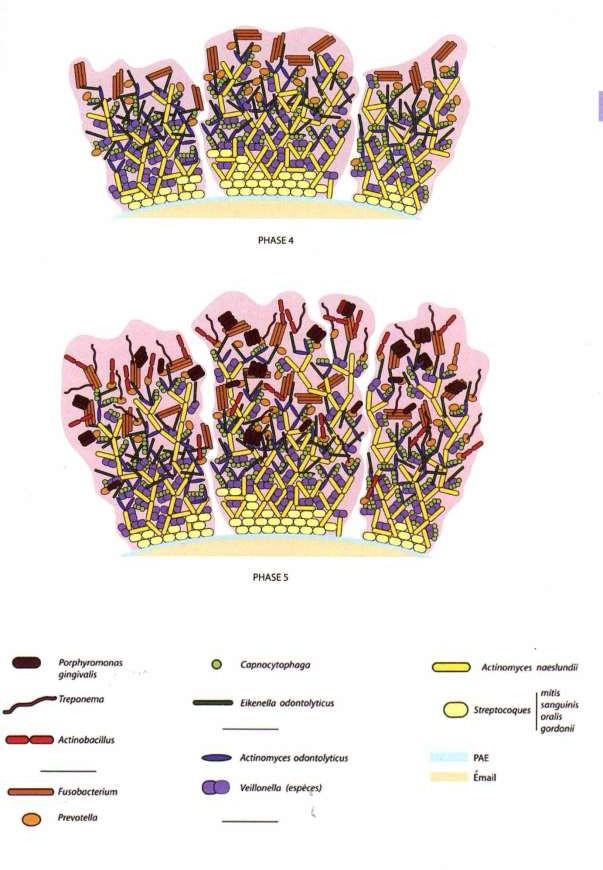
Sokransky’s bacterial complexes:
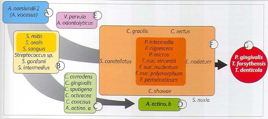
Schematic representation of the composition of bacterial complexes and their organization within a biofilm associated with periodontitis (Socransky and Haffajee)
Summary table of the main germs found in the oral ecosystem:
| Cocci gram (+) – Streptococcus- Peptostreptococcus | Cocci gram(-) -Veillonella-Neisseria |
Gram(+) bacillus -Actinomyces- Bifidobacterium-Eubacterium-lactobacillus-corynebacterium-propionibacterium | Bacillus gram( – ) – Actinobacillus- Bacteriodes-Compylobacter-Capnocytohphaga-Eikenella-Fusobacterium-Heamophilus-Porphyromonas-Prevotella-Selenomonas-Treponema |
VII-CONCLUSION:
Given the importance of the means of adhesion of germs of the oral flora , a simple cleaning of the mouth is not enough to eliminate it or to prevent its formation, hence the importance of using effective means of oral hygiene (mechanical brushing, brushing adjuvants: interdental floss, single-tuft brush, interdental brush, etc.
Oral Flora
Impacted wisdom teeth may require surgery.
Zirconia crowns are durable and aesthetic.
Bleeding gums may indicate periodontitis.
Invisible orthodontic treatments are gaining popularity.
Invisible orthodontic treatments are gaining popularity.
Modern dental fillings are both durable and discreet.
Interdental brushes are ideal for narrow spaces.
Good dental hygiene reduces the risk of cardiovascular disease.