COLLAGEN Periodontology Module
I-INTRODUCTION:
Collagen is the most abundant protein substance in the body (30% to 35% of the body’s proteins). It is the essential component of the extracellular matrix of connective tissue.
This glycoprotein is a component of all dental tissues except enamel and therefore plays an important role in the development, structure and physiology of the tooth and its supporting tissues.
It is part of two types of connective tissue:
-soft tissues: pulp, periodontium, gingival corium
-mineralized tissues: dentin, cementum, alveolar bone.
II-DEFINITION:
Collagen is an extracellular fibrous glycoprotein whose structural unit is TROPOCOLLAGEN . Collagen is organized into insoluble fibers that are very resistant to tension. This explains why it is the main constituent that provides strength to connective tissues such as bones, teeth, cartilage, tendons, ligaments, and the fibrous matrices of the skin and blood vessels.
It is also found in the extracellular matrix of connective tissue.
III-TROPOCOLLAGEN:
Tropocollagen is a triple helix of 03 polypeptide chains called alpha α with a length of 240-300nm and a diameter of 1.5nm; its molecular weight is 300000
Each alpha chain is made up of a sequence of repeating amino acids (Gly-ab) :
-glycine always present in every 2 or 3 amino acids,
-the a which is frequently a proline,
-the b a hydroxyproline.
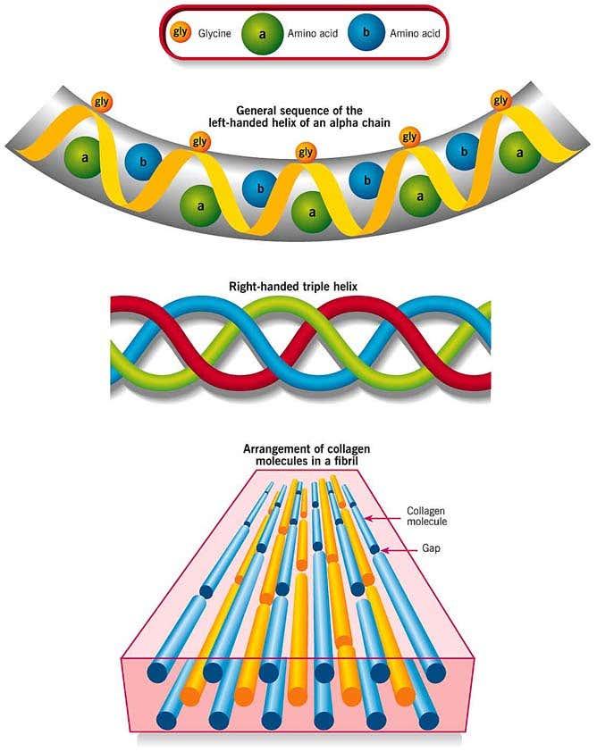
The number of amino acids making up one of these alpha chains is estimated at 1050, with more than 1000 being organized according to a repetitive triplet, except for the amino acids making up the two ends of the chain or telopeptides.
It is noted that it lacks tryptophan, which distinguishes it from serum proteins, and has few sulfur amino acids, which distinguishes it from keratins.
IV-THE DIFFERENT TYPES OF COLLAGEN:
There are at least 16 different types of collagen in the human body, the most common of which are collagen types (I II III IV V).
Depending on the distribution of amino acids, we distinguish 5 types of α chains :
– 4 chains called α 1 (from α 1 type 1 to α 1 type 4);
– a chain called α 2.
Depending on the distribution of these 5 types of chain, within the tropocollagen, we distinguish the types of collagen that characterize each connective tissue.
Collagens with identical α chains are said to be homotrimeric .
Collagen composed of two different chains is called heterotrimeric.

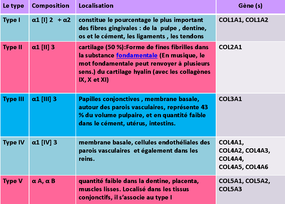
V. Distribution in dental tissues:
Collagen is an essential constituent of connective tissue, it is a component of all dental tissues except enamel, and it plays an important role in the development, structure and physiology of the tooth and its supporting tissues.
Collagen types I and III are mainly found.
Type I collagen is 7 times more than type III.
Type III is located at the papillae, around the vessels and under the basement membrane.
Type IV collagen is the major constituent of the lamina densa of basement membranes.
-At the level of the alveolo-dental ligament: type I collagen (approximately 80%), and type III (approximately 15%)
-At the alveolar bone level, type I collagen represents 90% of the organic phase of the bone.
-At the level of the cement represents 50 to 55%; it is found in the form of fibrils of 02 types:
*Intrinsic fibers: synthesized by cementoblasts
*Extrinsic fibers: (Sharpey fibers)
VI-THE BIO-SYNTHESIS OF COLLAGEN:
Collagen is synthesized by cells of natural origin
mesenchymal (stem, stromal and multipotent).
These cells can differentiate in a specific way
(odontoblasts in the pulp, osteoblasts in the bone tissue,
cementoblasts in cellular cementum and fibroblast in
connective tissue).
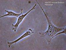
The key cell is the fibroblast : most often a polymorphic cell
fusiform or stellate, rich in organelles (rough reticulum,
golgi apparatus….) the cell membrane has folds
and invaginations where the presence of fibrils can be observed
of newly secreted collagen.
Fibroblasts are also capable of secreting many other
molecules of the ground substance (cytokines, growth factors, enzymes, elastin fibers, glycosaminoglycans, proteoglycans, etc.) and play an important role in tissue healing processes or in maintaining inflammatory reactions.
Collagen synthesis involves two steps, one intracellular while the other is extracellular.
A- THE INTRACELLULAR STAGE:
It is carried out at the level of:
-Granular endoplasmic reticulum (GER).
-From the Golgi apparatus (GA).
-Exocytosis vesicles (from the AG).
- At the level of the granular endoplasmic reticulum:
1. Synthesis of procollagen:
This is the ribosomal time: after the transcription of the DNA to result in the formation of a messenger RNA chain which will code the synthesis of collagen, the 02 parts of the ribosome come to rest on the messenger RNA to allow translation; the RNA transferase will thus come to place the amino acids which correspond to the coding of the messenger RNA chain to form the primary protein structure: a straight polypeptide chain.
All three alpha chains are synthesized at the same time
These chains have additional polypeptides at the N and COOH termini, or ‘pro’ regions.
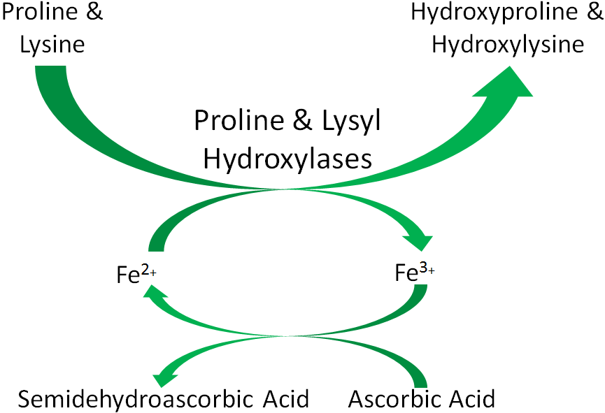
2. Hydroxylation of procollagen:
Hydroxylation of proline and lysine leading to the formation of hydroxyproline and hydroxylysine.
Hydroxylation requires the presence of O2, iron and ascorbic acid (vitamin C).
- At the level of the Golgi apparatus:
3. Glycosylation:
Saccharidic units (galactose or glycosylgalactose) are grafted onto the hydroxy of certain hydroxylysines, thanks to a transfer from UDP-galactose and UDP-glucose.
This step lasts 4 minutes with the presence of magnesium.
4. Formation of the propeller:
Once released from the ribosomes, the three alpha chains of procollagen are aligned in parallel and coil into a helix forming protropocollagen.
This association is initiated and stabilized by disulfide bond formation.
- At the level of exocytosis vesicles: Extrusion of tropocollagen
Once in contact with the cytoplasmic membrane, the exocytosis vesicles open onto the extracellular medium, and the protropocollagen molecule will transform into a tropocollagen molecule (cleavage) thanks to the procollagen peptidase which cuts the ends of the molecule (the coordination peptides).
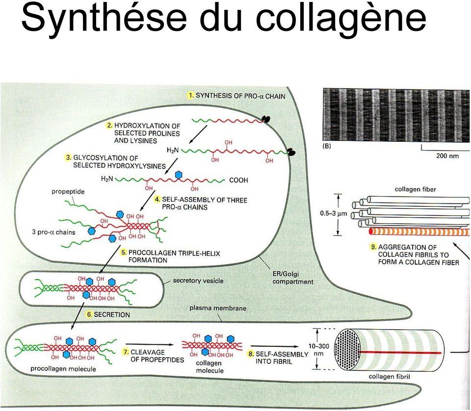
B- The extracellular stage or maturation of collagen:
Which will result in the formation of collagen fibrils by:
- cleavage of the pro regions: First the cleavage of the ‘pro’ regions thanks to procollagen-peptidases, extracellular proteases, thus releasing the tropocollagen.
- Creation of aldehyde functions: This is done through the oxidative deamination of lysine and hydroxylysine, under the influence of a lysyl-oxidase.
- Polymerization: Creation of intermolecular bridges between these neighboring tropocollagen aldehyde functions to allow their binding
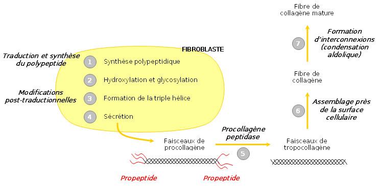
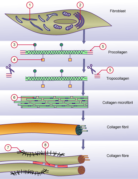
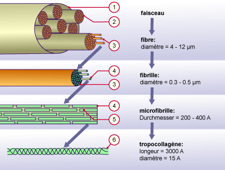
Formation of collagen fiber
VI. Regulation of collagen production:
Although much remains to be discovered about the mechanisms of transcriptional regulation, certain molecules are known to modify the level of expression of collagens:
- TGF-β (Transforming Growth Factor) increases the transcription of genes corresponding to extracellular matrix proteins, but also makes collagen mRNA more stable,
- Interferon γ (IFN-γ) is a cytokine produced by lymphocytes. IFN-γ decreases both fibroblast proliferation and type I collagen synthesis by these cells.
- TNF-α (Tumor necrosis factor α) and interleukin-1 are two other cytokines regulating collagen production and both are produced by macrophages. Although their mechanisms of action are different, their results are similar: they stimulate fibroblast proliferation, but inhibit type I collagen production and increase the production of interstitial collagenases and therefore collagen degradation.
VII- Collagen degradation:
A- Phagocytic mechanism:
This is the mechanism by which the cell captures a solid element to digest it. The phagocytic cell generally has a very mobile membrane forming hyaluroplasmic veils and possibly capable of liquefying to surround the particle.
Phagocytosis is carried out by several cells with phagocytic activity: fibroblasts with degradative activity, osteoclasts, and cementoclasts.
This phagocytosis takes place in the following three (3) stages:
-Adhesion and Capture: after attraction and adhesion of the foreign body with the cell membrane.
– Engulfment (or ingestion phase): the product is engulfed in the cell inside a vacuole called a phagosome.
-Digestion (or intracytoplasmic destruction phase): The fusion of the phagosome with lysosomes (enzymes) is at the origin of the Phagolysosome where the destruction of the foreign body will take place.
B-Enzymatic mechanism:
It is carried out by proteolytic enzymes, for example tissue collagenases which can be synthesized by several cells with macrophagic activity: fibroblast with degradative activity, osteoclast, and cementoclast…..
Tissue collagenase is found in the extracellular environment in an inactive form (procollagenase) and under the influence of activators (cytokines, bacteria, etc.), it transforms into true active tissue collagenase so that inhibitors (serum proteins, estrogen hormone, etc.) can inactivate it again.
The regulation of collagenolytic activity depends on: the type of collagen itself (type II more resistant than type I), age, variations in temperature and pH, and certain proteins and hormones.
VII- Physiology of collagen:
1-Collagen is part of the extracellular matrix: it is responsible for the cohesion of tissues on the one hand; and also gives resistance, flexibility and elasticity to the different tissues on the other hand.
2- resistance and firmness: Collagen resists traction, and it is essential for the healing process.
3-structural protein: It is involved in the formation of: skin, tendons, bones, especially: Type I (for example).
4-Turn over collagen:
It is the balance between synthesized collagen and degraded collagen, of which the fibroblast is the key cell.
The renewal of desmodontal collagen fibers allows the control of dental micromovements.
The collagen of the desmodont has a turnover 5 times faster than that of the gingival corium and the alveolar bone and 15 times faster than the dermis, however this turnover slows down with age.
5-mineralization: The presence of collagen is essential for mineralization.
6-the eruption: By the game of collagen renewal: the newly synthesized molecule is longer; this process creates tension forces which cause the movement of the tooth and its eruption.
X-CONCLUSION:
Collagen is an essential constituent of connective tissues , it is part of the composition of all periodontal tissues and plays a major role in periodontal repair and regeneration . Its alteration will affect the health of the periodontium.
COLLAGEN Periodontology Module
Impacted wisdom teeth may require surgery.
Zirconia crowns are durable and aesthetic.
Bleeding gums may indicate periodontitis.
Invisible orthodontic treatments are gaining popularity.
Invisible orthodontic treatments are gaining popularity.
Modern dental fillings are both durable and discreet.
Interdental brushes are ideal for narrow spaces.
Good dental hygiene reduces the risk of cardiovascular disease.