The first lower premolar
- Timeline:
- Start of calcification: 21 to 24 months.
- End of coronary calcification: 5 to 6 years.
- Eruption: 9 to 12 years.
- End of apical calcification: 12 to 13 years .
- Average measurements:
Total height: 22.5 mm.
Crown height: 8.5mm.
Coronal mesio-distal diameter: 7 mm.
Coronal vestibulo-lingual diameter: 7.5 mm
- Description
- Vestibular face:
- The crown:
- The general outline is that of a pentagon like
for the canine.
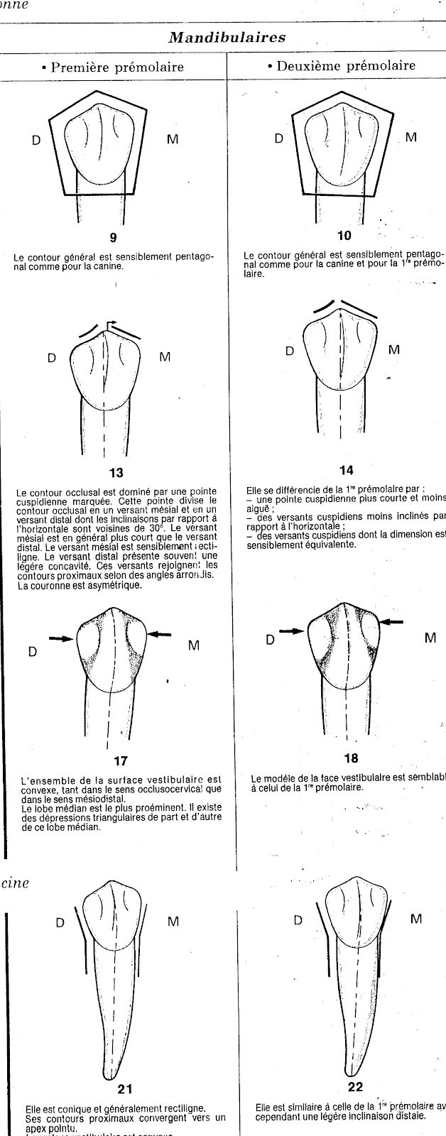
- The occlusal contour is dominated by a point
marked cusp. This point divides the outline
occlusal in 2 slopes: one mesial and the other distal.
- The mesial slope is generally short and
straight while the distal slope is long and
concave. These slopes join the contours
proximal with rounded angles.
- The crown is asymmetrical.
- The entire vestibular surface is
convex, both in the occluso-cervical direction
than in the mesiodistal direction.
- The median lobe is the most prominent, there are
triangular depressions on either side of
this median lobe.
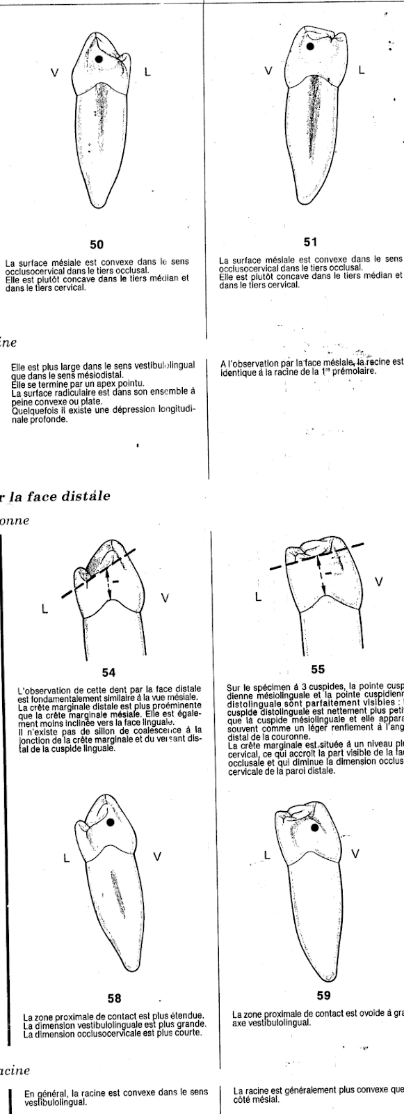
- The root:
- It is conical and generally rectilinear.
- Its proximal contours converge towards a
pointed apex.
- The vestibular surface is convex.
- Lingual face:
- The crown :
- The entire lingual surface is smaller than the face
vestibular.
- More than half of the occlusal area is visible at
observation by this face: this is due to the strong
lingual inclination of the occlusal surface.
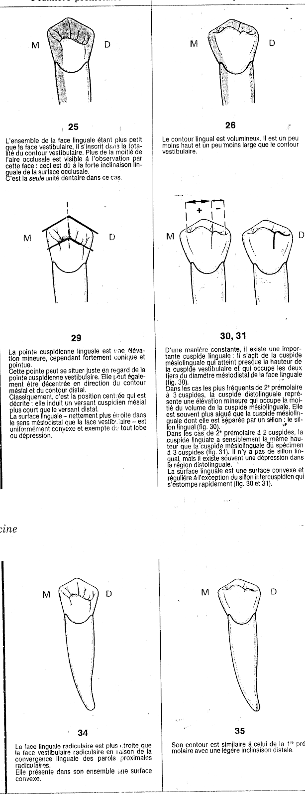
- The lingual cusp tip is a minor elevation,
However, it is strongly conical and pointed, it can
occupy a middle position as it can be
offset to the mesial or distal side.
- The lingual surface is uniformly convex and free
of any lobe or depression.
- The root:
- The root lingual surface is narrower
that the root vestibular face due to
of lingual convergence of the proximal walls
root
- It presents as a whole a surface
Convex.
- Mesial face:
- The crown:
- The general outline is trapezoidal; this is linked to a
very short lingual contour.
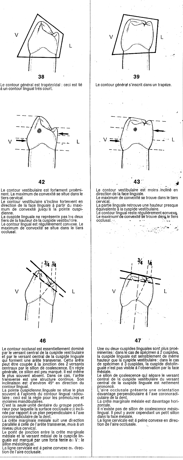
- The vestibular contour is strongly prominent
and the maximum convexity is located in the 1/3
cervical, from this 1/3, the vestibular contour
tilts sharply in a lingual direction to the
cusp tip.
- The lingual contour is convex and the maximum
convexity is located in the occlusal third.
- The lingual cusp tip is most often located
in line with the root lingual contour: this is a
rule for mandibular premolars.
- It is the only dental unit in the posterior group for
which the occlusal surface is inclined relative to
the coronoradicular axis of the tooth.
- The cervical line is barely convex towards
the occlusal area
- The mesial surface is convex in the direction
occlusocervical in the occlusal 1/3. It is rather
concave in the medial 1/3 and in the cervical 1/3
- The maximum convexity of the mesial wall is located
at the junction of the vestibular 1/3 and the medial 1/3
in the vestibulo-lingual direction and at the junction of 1/3
occlusalet of the median 1/3 in the occluso-cervical direction
- The root:
- It is wider in the vestibulolingual direction than
in the mesiodistal direction
- It ends with a pointed apex
- The root surface as a whole is barely
convex or flat
- Sometimes there is a longitudinal depression
deep.
- Distal face:
a. the crown:
- Observation of this tooth from the distal face is
basically similar to the mesial view.
- The distal marginal ridge is more prominent than
the mesial marginal ridge.
- The proximal contact zone is larger.
b.the root:
- In general, the root is convex in the
vestibulolingual sense
- Occlusal face:
- The general outline resembles that of a
” cut diamond ”
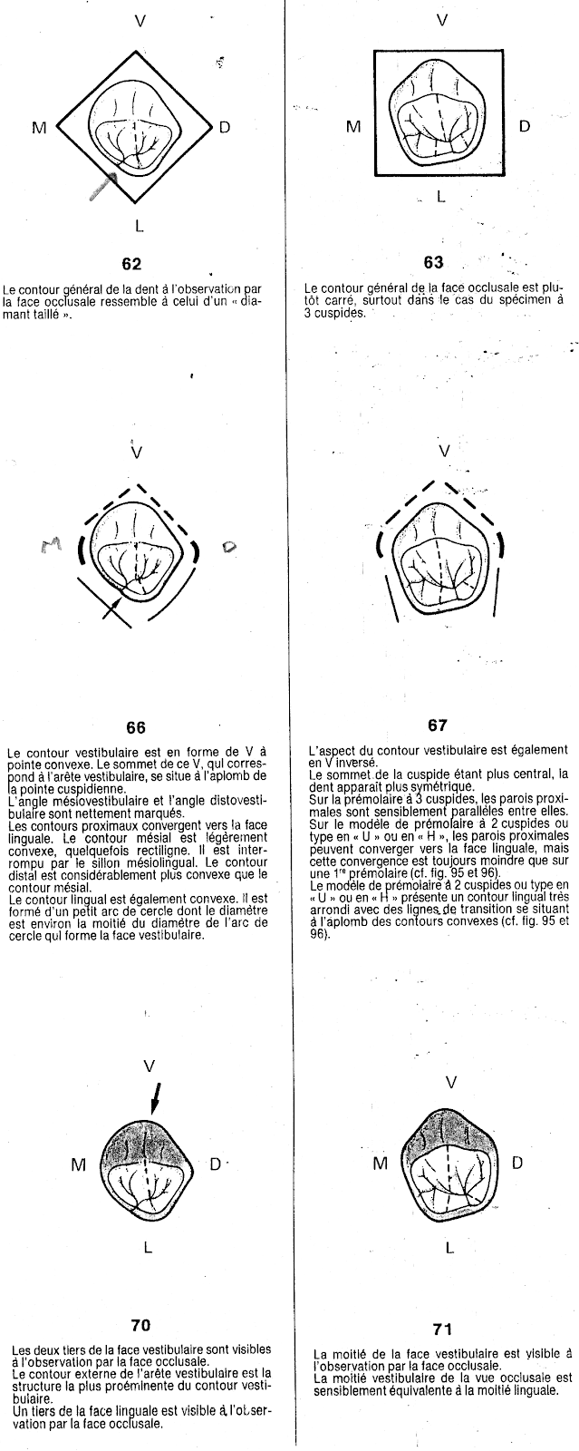
- The vestibular contour is “V” shaped
with a convex tip, the summit of this V, corresponds to
the vestibular ridge, and is located directly above the tip
cusp.
- Proximal contours converge toward the face
lingual, the mesial contour is slightly convex
sometimes straight it is interrupted by the furrow
mesiolingual the distal contour is considerably
more convex than the mesial contour
- The lingual contour is convex. It is formed by a small circular arc whose diameter is approximately half the diameter of the circular arc that forms the vestibular surface.
- The occlusal area is triangular: the base of the triangle
corresponds to the mesial and distal slope of the cusp
vestibular, and the summit corresponds to the tip of the
lingual cusp.
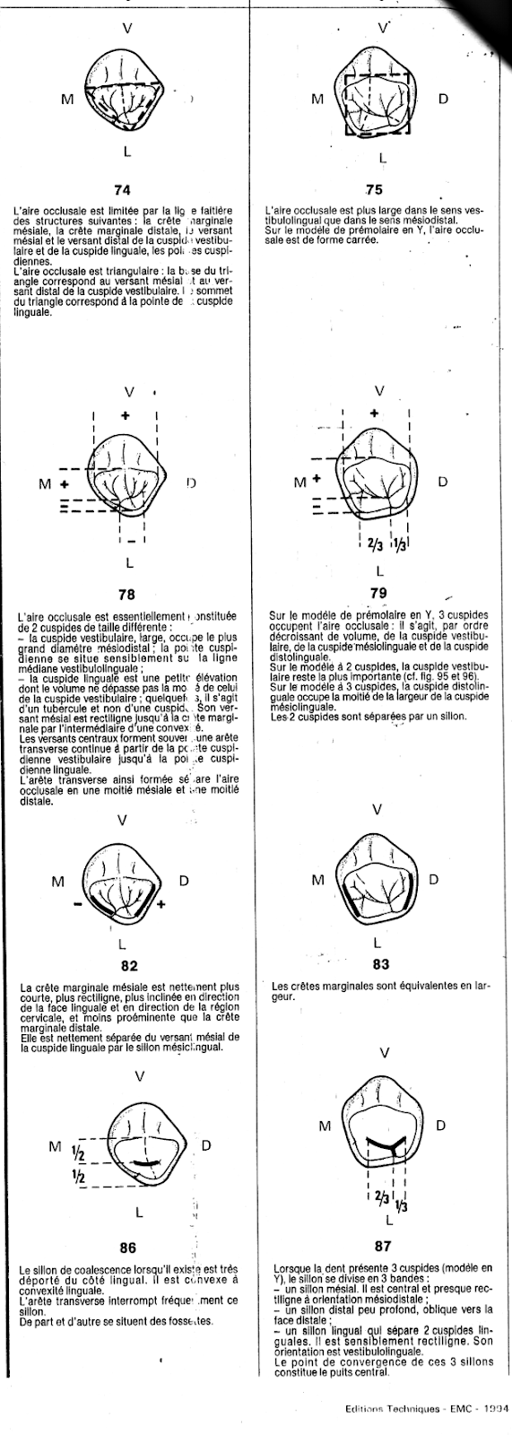
- The occlusal area is made up of 2 cusps of
different size:
* The wide vestibular cusp occupies the most
large mesiodistal diameter, the tip
cusp is located approximately on the line
median vestibulolingual.
* The lingual cusp is a small elevation
whose volume does not exceed half of
that of the vestibular cusp.
- The mesial marginal crest is distinctly shorter,
more straight, more inclined towards the region
cervical, and less prominent than the marginal crest
distal.
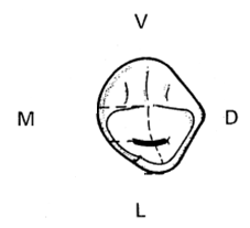
- The central slopes of these cusps often form
a continuous transverse edge from the tip
vestibular cusp to cusp tip
lingual, this ridge separates the occlusal area into one half
mesial and one distal half.
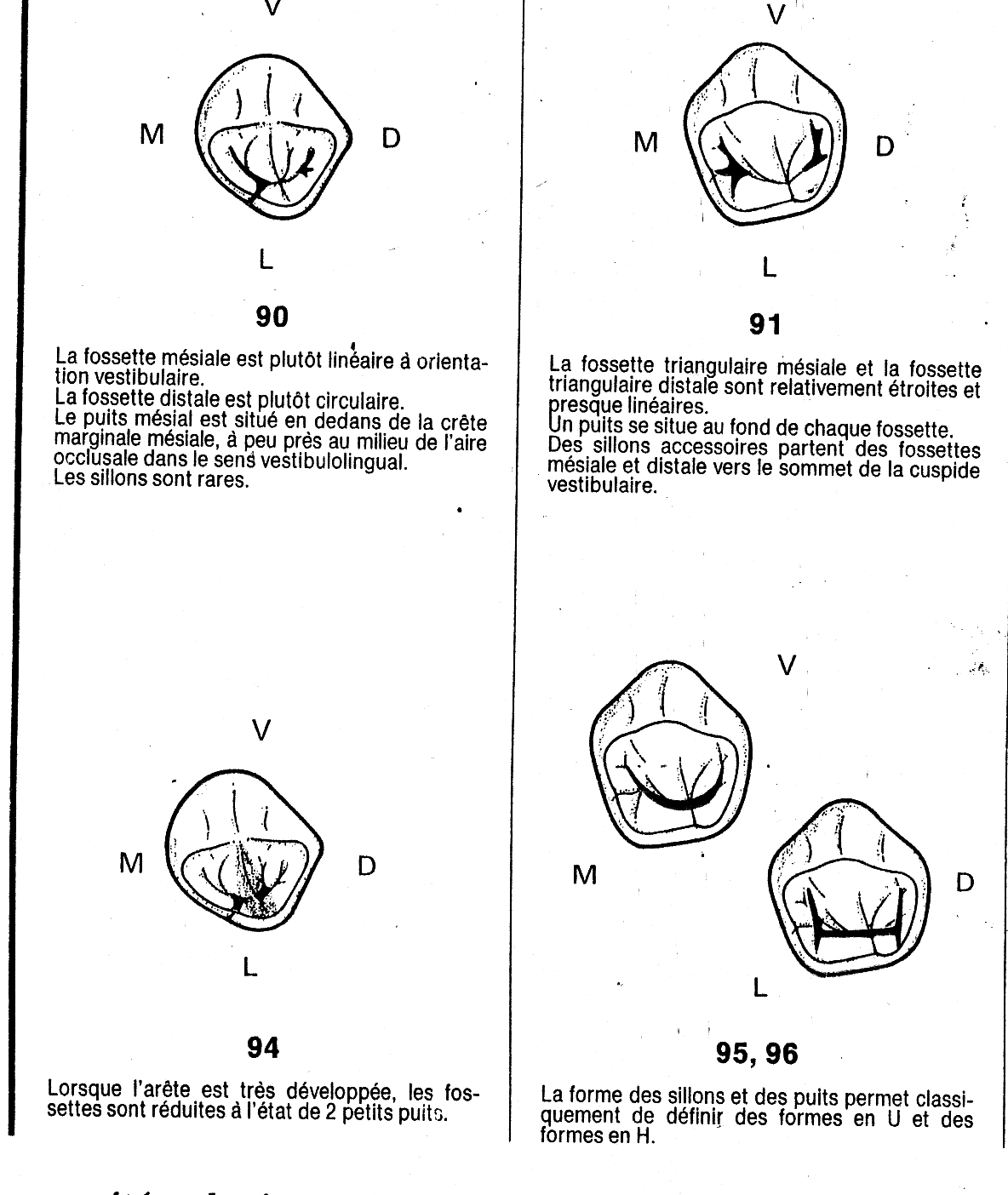
- The central groove , when it exists, is very far from the
On the lingual side, the transverse ridge often interrupts this
furrow. On either side are dimples .
The first lower premolar
Impacted wisdom teeth may require surgery.
Zirconia crowns are durable and aesthetic.
Bleeding gums may indicate periodontitis.
Invisible orthodontic treatments are gaining popularity.
Invisible orthodontic treatments are gaining popularity.
Modern dental fillings are both durable and discreet.
Interdental brushes are ideal for narrow spaces.
Good dental hygiene reduces the risk of cardiovascular disease.