The second upper premolar
- Timeline:
- Start of calcification: 24-27 months
- End of coronary calcification: 6-7 years
- Eruption: 10-12 years
- End of apical calcification: 12-14 years
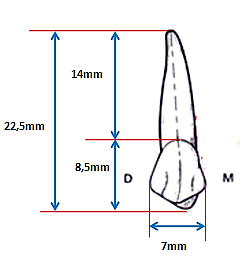
- Average measurement:
- Total height = 22.5 mm
- Crown height = 08.5 mm
- Coronal mesial-distal diameter = 7mm
- Coronal vestibulo-lingual diameter = 9mm
- Description
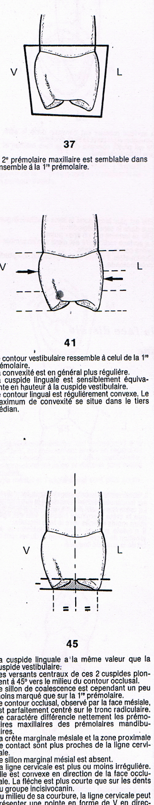
- Vestibular face
- The crown
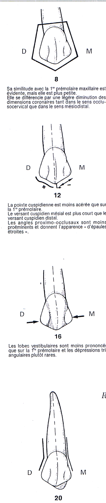
- Its similarity to the upper 1st premolar is
obvious but it is smaller.
- The cusp tip is less
sharper than the upper 1st premolar .
- The mesial cusp slope is shorter than the
distal slope.
- The proximal angles are less prominent and
give the appearance of “narrow shoulders.”
- The vestibular lobes are less pronounced and
depressions are rare.
- The root:
- The 2nd premolar generally has only one
Root.
- It is a little longer and a little narrower than
that of the 1st premolar .
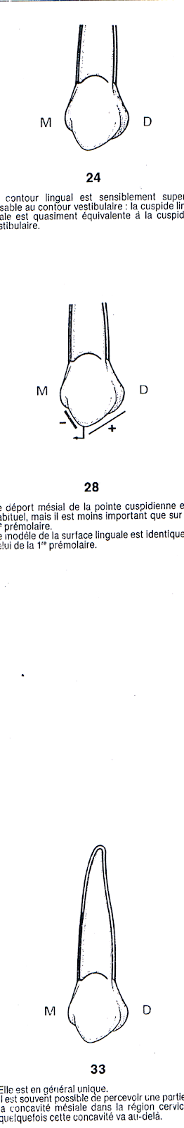
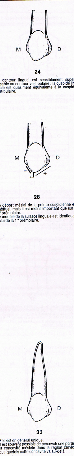
- Palatal face:
- The crown
- Palatal contour is substantially superimposable on the vestibular contour (the palatal cusp is almost equivalent to the vestibular cusp).
- The mesial offset of the cusp tip is less significant than on the 1st premolar .
- The root:
- It is generally unique
- It sometimes has two apices
- Mesial face
- The crown
- The 2nd premolar is generally similar to the 1st premolar .
- The vestibular outline resembles that of the 1st premolar .
Convexity is generally more regular; maximum convexity is between the junction of the cervical 1/3 and the medial 1/3 or in the medial 1/3
- The palatal cusp has the same value as the vestibular cusp.
- The palatal contour is regularly convex, the maximum convexity is located in the middle 1/3
- The occlusal contour observed by the mesial face is perfectly centered on the root trunk.
- The lingual cusp is approximately the same height as the vestibular cusp.
- The mesial marginal groove is absent,
- The cervical line is more or less irregular
- The root
- Distal face
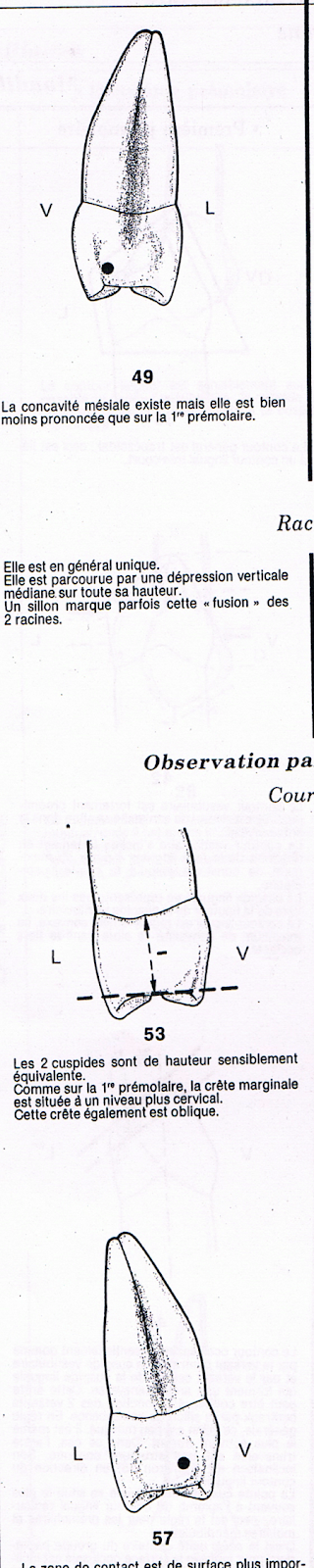
b. The root:
- It is generally unique
- It is crossed by a vertical depression
median over its entire height
3.4 Distal face:
- The crown :
- The two cusps are of height
equivalent, the marginal ridge is located at
a more cervical level and it is oblique.
- The contact area is of larger surface area
important.
- The distal face appears more convex.
- The root:
- It presents a vertical depression
less marked than on the mesial side
- Occlusal face
- The crown
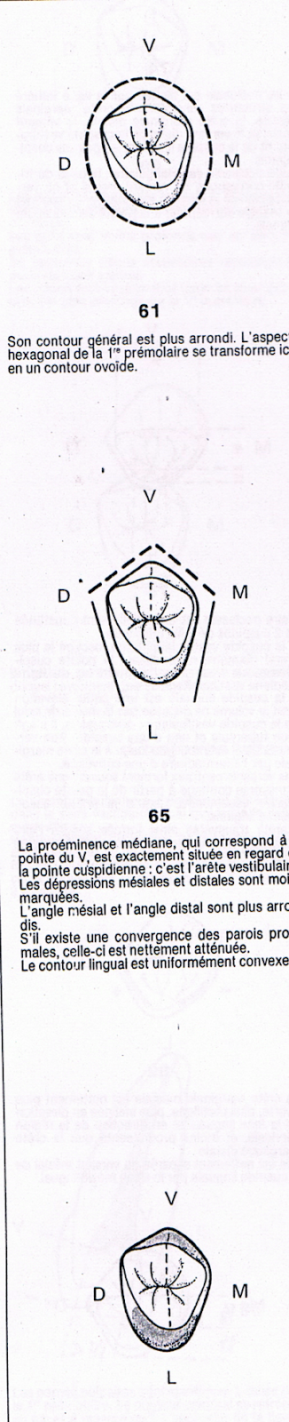
- The 2nd premolar has an ovoid outline.
- The median prominence, which corresponds to the
tip of the V, is located exactly opposite
of the cusp tip: it is the edge
vestibular.
- The mesial and distal corners are rounded.
- The palatal contour is convex.
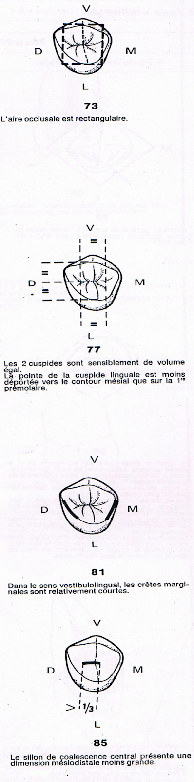
- The two cusps are substantially of volume
equal
- The tip of the palatal cusp is less offset
towards the mesial contour than on the first premolar.
- The marginal ridges are relatively short
- The central groove has a mesio-distal dimension
less large.
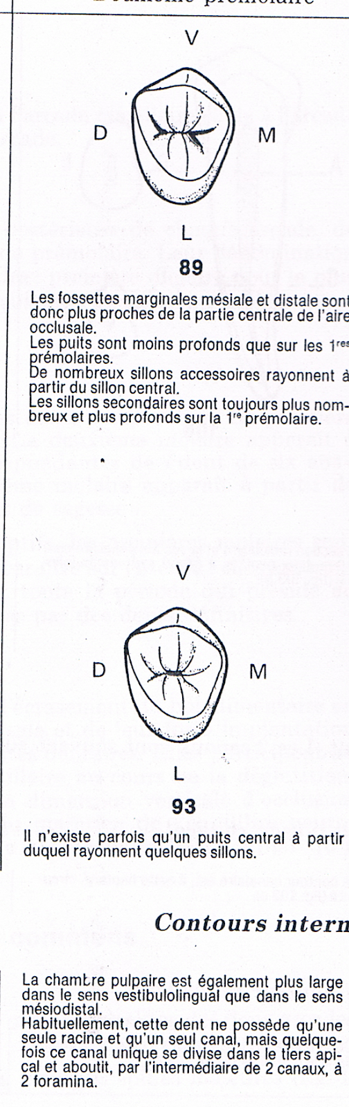
- Numerous accessory furrows radiate from the
central furrow
- These accessory furrows are always more numerous and
deeper on the 1st premolar
4. Endodontic anatomy:
The pulp chamber is also wider in the vestibulo-lingual direction than in the mesio-distal direction.
Usually, this tooth has only one root and one canal, but sometimes this single canal divides in the apical third and ends, through 2 canals, in 2 foramina.
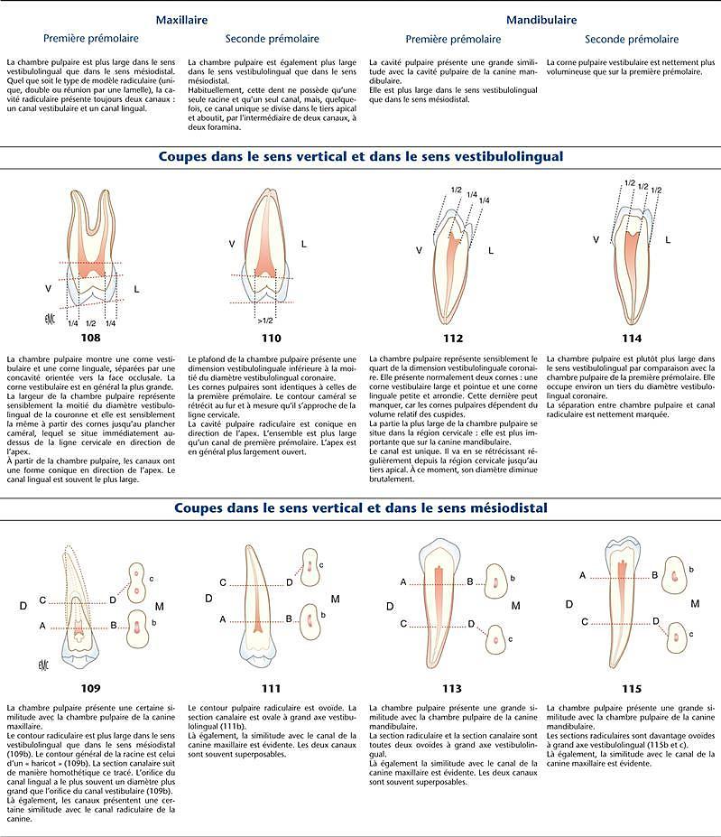
Vestibulo-lingual sense
The pulp chamber:
The pulp horns are identical to those of the 1st premolar . The cameral contour narrows as it approaches the cervical line.
Root canals:
The root cavity is conical towards the apex. The whole is wider than a 1st premolar canal . The apex is more widely open.
4.2. Horizontal sections:
The outline of the pulp cavity is superimposable on the general outline of the pulp cavity of the canine or the 1st premolar
In 15% of cases this tooth has 2 distinct roots, each with a canal.
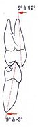
5. SITUATION IN THE MOUTH:
5.1. Vestibulo-lingual direction : The vestibular face of the crown is vertical. The lingual cusp descends slightly lower than the vestibular cusp. The root is inclined on the lingual side (approximately 17° from the vertical).
5.2. Mesio-distal direction : Crown and root direction parallel to that of the upper first premolar.
The second upper premolar
Impacted wisdom teeth may require surgery.
Zirconia crowns are durable and aesthetic.
Bleeding gums may indicate periodontitis.
Invisible orthodontic treatments are gaining popularity.
Invisible orthodontic treatments are gaining popularity.
Modern dental fillings are both durable and discreet.
Interdental brushes are ideal for narrow spaces.
Good dental hygiene reduces the risk of cardiovascular disease.