Instrumentation in Endodontics
Introduction
In the majority of cases, endodontic therapy consists of eliminating the intra-canal contents, optimally disinfecting the endodontic network and then sealing it in a three-dimensional, watertight and long-lasting manner.
To meet these objectives it requires the manual or mechanical use of various instruments.
Classification of endodontic instruments
1. Instruments for exploration and diagnosis in endodontics
2. Instruments for dental isolation (the operating field)
3. Instruments required for endodontic access cavity
4. Root canal shaping instruments
5. Instruments for root canal obturation
1. Instruments for exploration and diagnosis in endodontics
2. Instruments for dental isolation (the operating field)
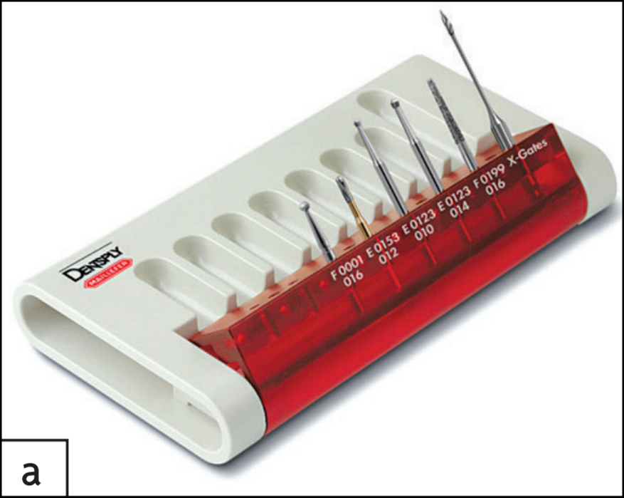
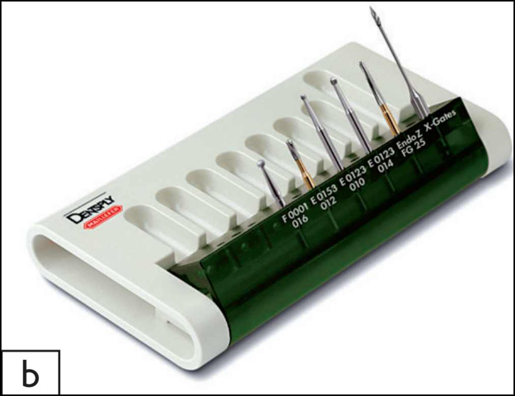
3. Instruments required for endodontic access cavity
• A 016 diameter diamond ball bur. Its abrasive action facilitates the removal of enamel or ceramic on ceramic-metal or ceramic-ceramic copings;
• A transmetal bur that can pass through a metal crown or the infrastructure of ceramic-metal crowns;
• Two long-necked tungsten carbide ball burs. The long neck allows for clear vision of the operating field under optical aid (operating microscope or loupes):
– the one with a diameter of 010 is used for the mandibular incisors
– the one with a diameter of 014 is for all the other teeth;
• A 016 diamond round bur (Cavity Access® Set): Its grain size allows the cavity to be widened and smooth walls to be obtained during finishing. Its tip is active and must not act on the cavity floor;
• A Zekrya Endo bur (Cavity Access® Z Set). With an active blade, it allows you to widen and finish the cavity while avoiding iatrogenic action with its blunt tip;
• An X-Gates drill, and instrument allows marking of the coronal orifices by pointing the canal entrance and performing a brushing movement on withdrawal against the wall. This drill corresponds to the combination of conventional Gates drills No. 1, 2, 3 and 4. It should not be used as an intracanal shaping instrument
Instrumentation in Endodontics
•Ultrasonic instruments: are indicated for the discovery of hidden canal orifices and for finishing the walls of the cavity by eliminating any overhangs present.

Instrumentation in Endodontics
•Endoflare, nickel-titanium instrument for the removal of dentinal overhangs at canal entrances and the preparation of the coronal third of canals.
Instrumentation in Endodontics
•Hand instruments are used to locate coronal overhangs, remove cameral pulp, locate canal entries
Excavators for removing cameral pulp.
Exploratory probes with a small hook for locating overhangs (probe no. 17)
Straight endodontic probes longer than conventional probes to locate canal entrances (e.g. Rhein probe)
4. Root canal shaping instruments
4.1. Conditioning of instruments:
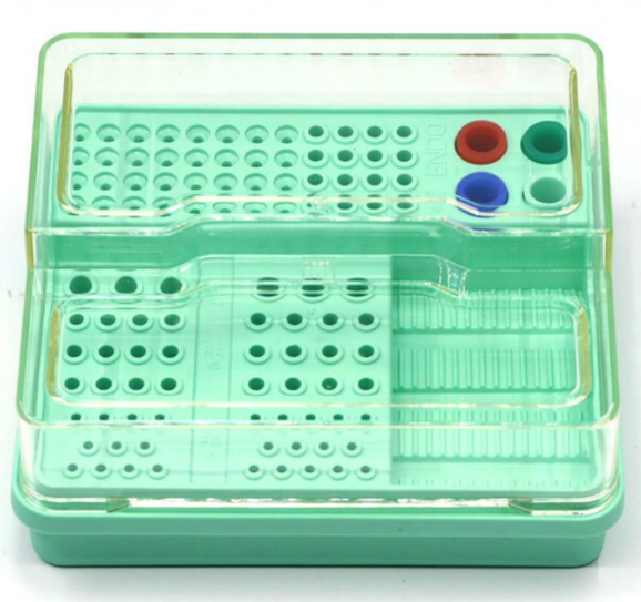
- Endodontics box
Allows you to store and arrange all the instrumentation necessary for endodontic procedures and to prepare an instrumental sequence
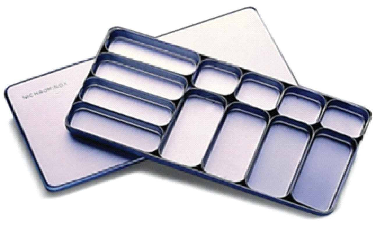
- Pulpectomy box
Made of aluminum, with rectangular interior compartments allowing the sterilization and storage of endodontic instruments
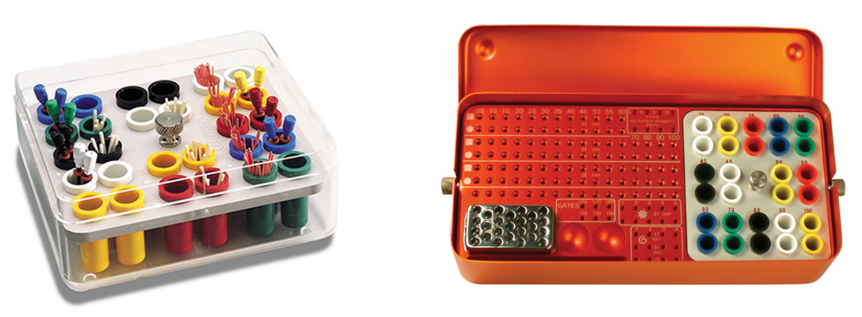
- Endobox
Small metal box for storing and sterilizing endo-canal tips and canal instruments
Instrumentation in Endodontics
- Transfer sponge :
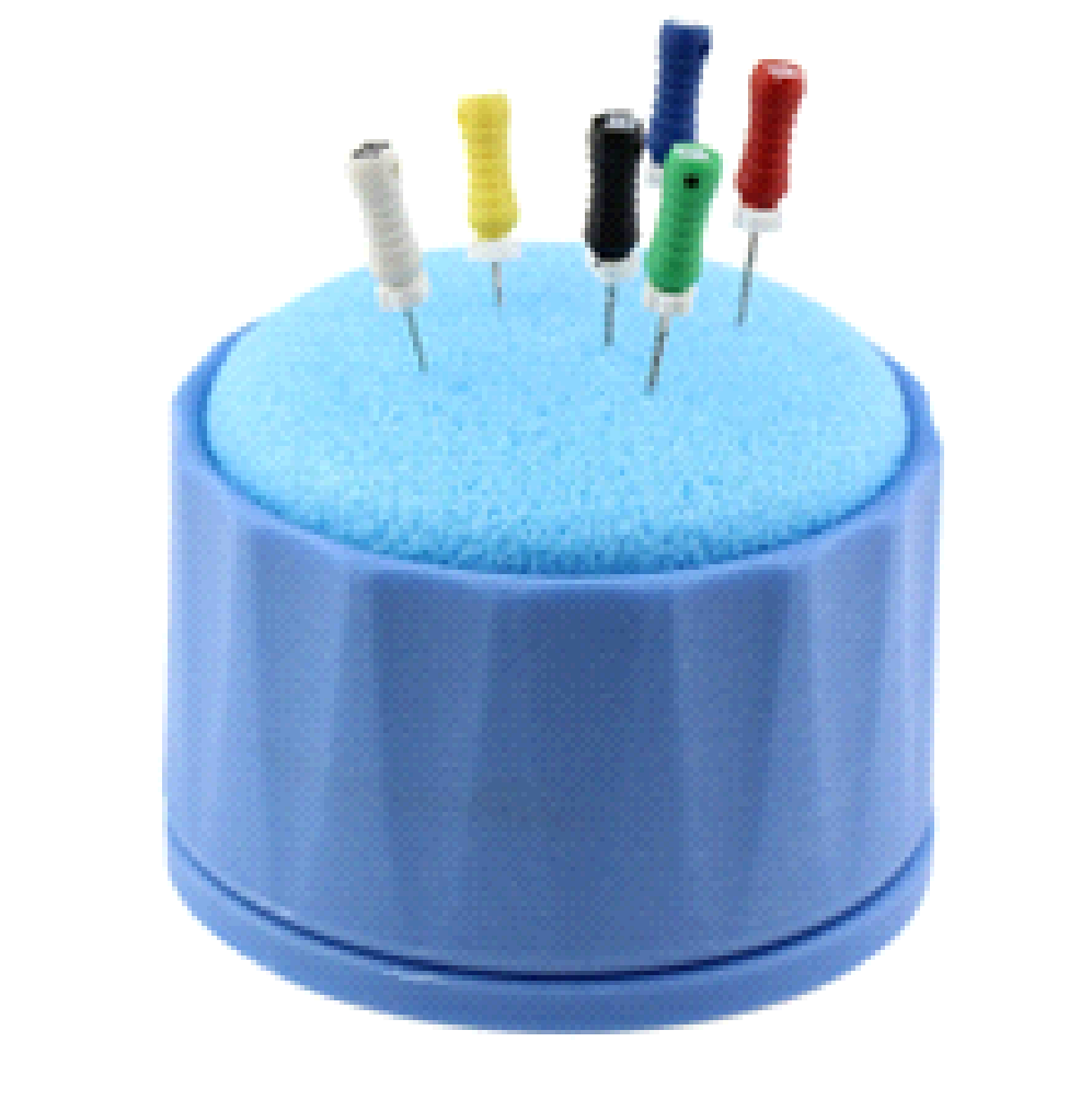
Facilitates endodontic work and avoids injections
4.2. Alloys used in the manufacture of endodontic instruments
– Endodontic instruments are made either:
- In stainless steel,
- Either in shape memory alloy based on nickel and titanium.
- Stainless steels
– Stainless steels are groups of iron-based metals containing at least 10% chromium.
– Those used for endodontic instruments contain 18% chromium. This chromium helps protect the iron from oxidation, hence the name “stainless” steel. - Nickel-Titanium (Ni-Ti) alloy
– These alloys are composed of 50% nickel and 50% titanium.
– The advantage of this alloy is its superelastic properties: the alloy has an elastic behavior that allows it to have a reversible deformation of up to 8%.
– Ni-Ti is therefore part of the shape memory alloys (SMA).
– It is this superelastic property as well as good mechanical properties that make this alloy so attractive for the design of dental instruments, particularly endodontic ones.
4.3. Stainless steel manual root canal preparation instruments
4.3.1. General description of an instrument

Instrumentation in Endodontics
4.3.2. Characteristics of ISO standard stainless steel manual root canal preparation instruments
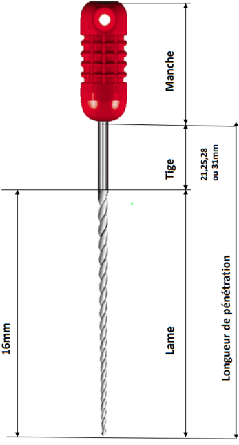
a. Instrumental length
The active blade has a fixed length of 16 mm, in addition, the total length of the instrument is available in 5 sizes: 19, 21, 25, 27 and 31 mm the longer an instrument is, the more flexible it is (with identical diameters and conicities)
b. Instrument diameters and color coding
We can define 2 reference diameters for the instrument: D0 and D16. These diameters are expressed in hundredths of a millimeter.
D0: dimension of the section of the instrument at 1 mm from the tip of the active blade. It is this diameter which corresponds to the number of the instrument, and therefore to its color.
D16: dimension of the section of the instrument at the other end of the active blade, i.e. 16 mm from D0.
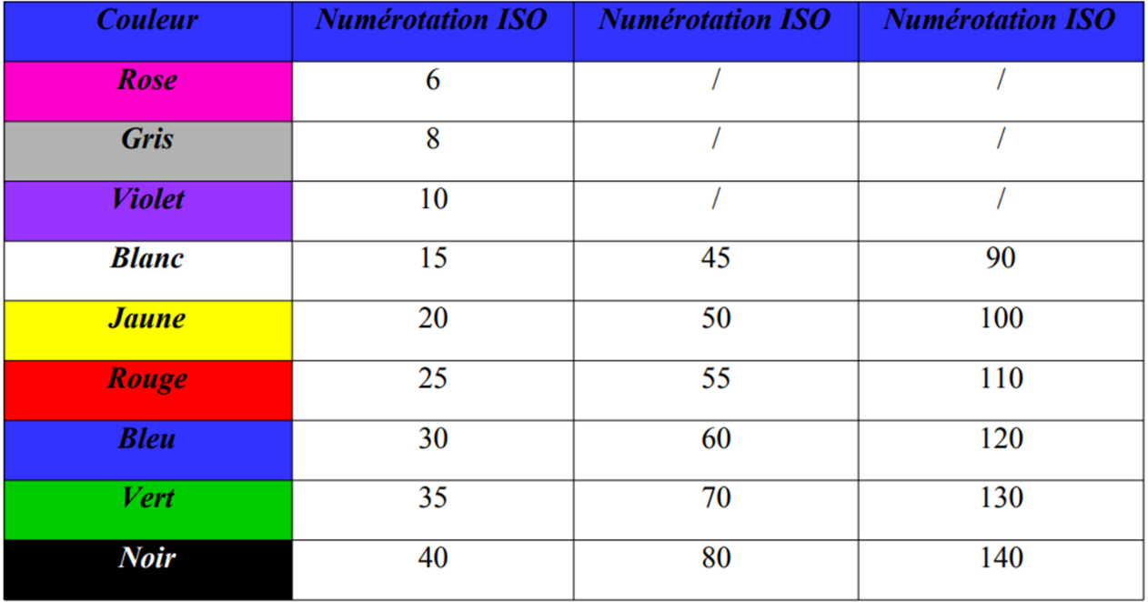
Color code and ISO (International Standard Organization) standard.
- From n°10 to n°60 the increase in diameter is made by 5/100 mm
- From n°60 to n°140 the increase in diameter is made by 10/100 mm
c. Conicity
Taper, on the other hand, corresponds to the increase in diameter per millimeter along an instrument or canal. Instruments can have the same diameter at the tip but different tapers. For a 30/100 diameter at the tip, the 2% taper instrument sees its diameter increase by 0.02 mm (2/100 of a mm) per millimeter of length; at 1 mm from the tip, its diameter will be 0.32 mm (32/100). On the 6% taper instrument, the diameter will be 0.36 mm (36/100) at 1 mm from the tip.
Taper and apical diameter for all stainless steel manual root canal preparation instruments complying with ISO standard, the taper is 2%
d. Manufacturing process
– Stainless steel root canal preparation instruments can be machined or twisted
e. Instrumental section
- There are 3 types of instrumental sections for stainless steel manual root canal preparation instruments: square, triangular or round.
- The instrumental section will influence the strength, flexibility and centering of the instrument in the canal.
Instrumentation in Endodontics
f. Helix angle
The helix angle is the angle formed between the axis of the instrument and the axis of the coils. It has an influence on the coronal evacuation of debris and on the screwing effect of the instrument in the canal in a more or less pronounced manner.
For ISO standard instruments, the helix angle can typically be 20° for broaches, 40° for K files, or 60° for H files.
The more closed this angle, the more active the instrument is in rotation; the more open the angle, the more effective the instrument is in traction.


Helix angles of an H file and a K file
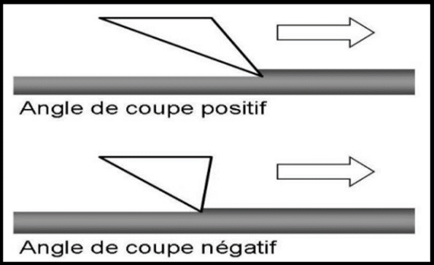
g. The cutting angle
The angle at which the blades approach the canal walls. It is directly associated with the cutting efficiency of the instruments.
This angle, also called the angle of attack, can be: – Positive, the cut is active. – Neutral, the cut is perpendicular to the wall of the canal. – Negative, the cut is passive, the instrument acts by smoothing.
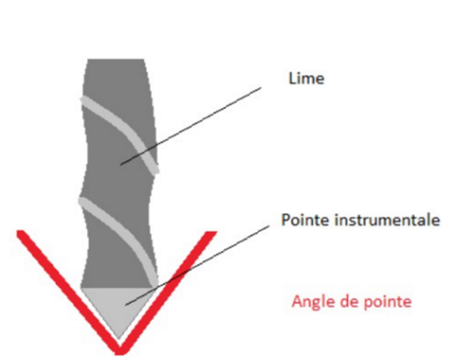
h. Point angle
The tip angle is formed by the intersection of the blades at the end of the instrument.
According to the ISO standard, it varies from 60 to 90°.
The tip angle can be passive, i.e. non-working: this is the case for most Ni-Ti endodontic instruments, which allows the instrument to be guided safely into the canal.
It can also be active root canal retreatment
Instrumentation in EndodonticsInstrumentation in Endodontics
Diagram of the tip angle (Dentalespace)
- No propeller
The helix pitch refers to the distance between two consecutive turns. For two instruments of the same diameter, section, alloy and taper, it will influence the flexibility of the instrument and its screwing effect when used in rotation.
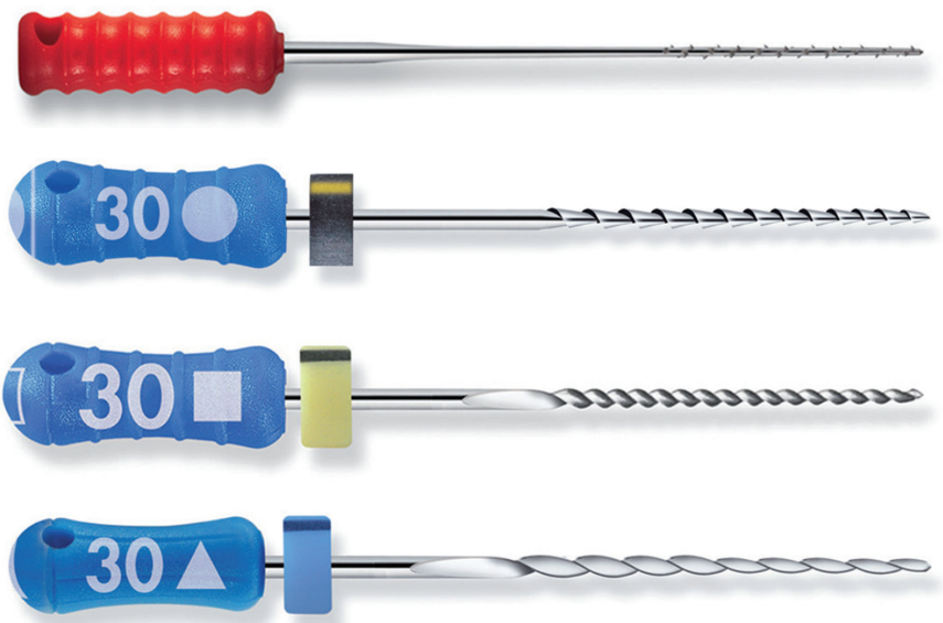
4.3.3. Main reference stainless steel hand instruments
a. Nerve puller
Consisting of a parallel body in which notches are made to create barbs, the nerve puller can be used occasionally to remove the pulp in one piece before root canal shaping. It can also be used to hook and remove paper points or cotton balls for example.

b. The files
- K (Kerr) files
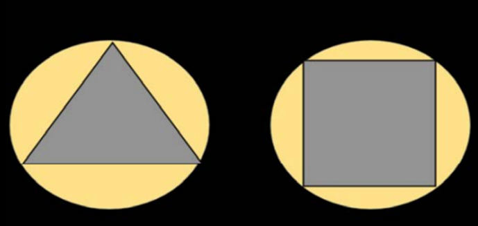
K files are instruments twisted from a square blank
The identification symbol is a square.
There are more turns than pins (1.5 to 2.5 turns per mm), so a larger helix angle of 40° on average.

The use of these files is mainly carried out in traction and/or rotation/traction
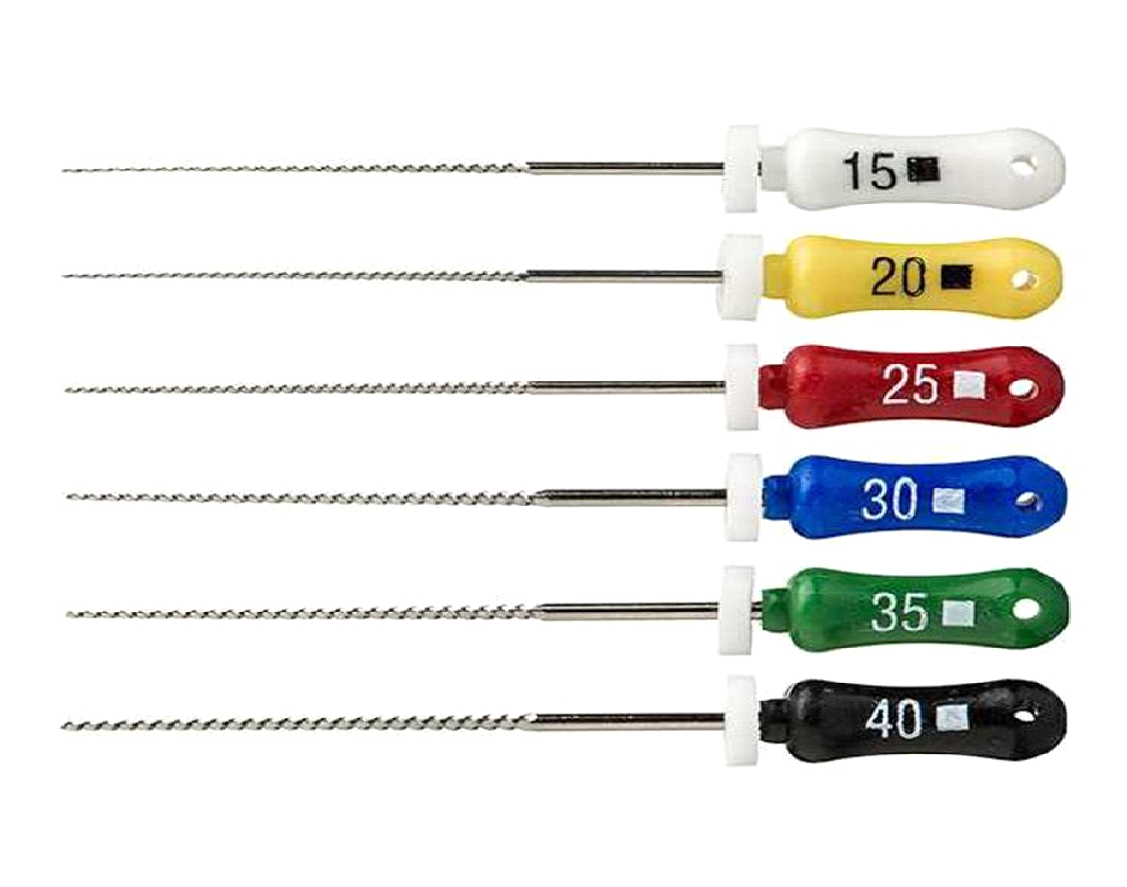
Files remain more rigid instruments than pins, therefore more effective in initial penetration.
Also used in the phases of spotting, permeabilization (or recapitulation) and widening.
- Flexofile

Derived from the K file
Diameters from 8/100th →80/100th mm,
Lengths (mm): 21, 25, 31,
Twisted, triangular section, used in rotation/traction,
Great flexibility,
Used during catheterization.

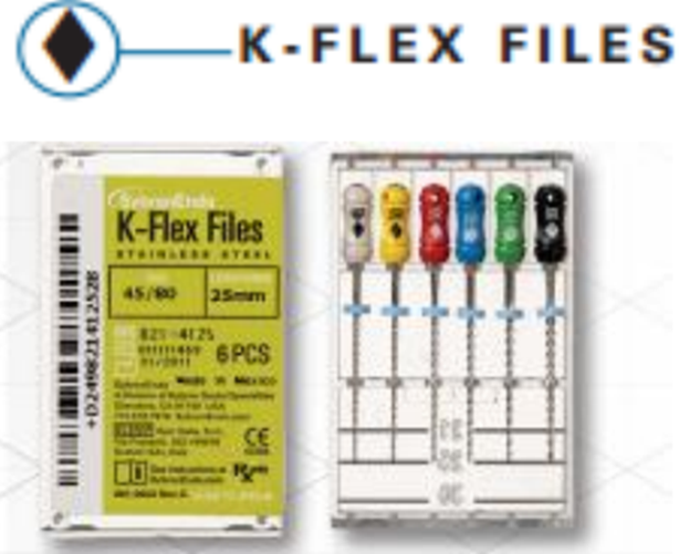
- The K.Flex
Twisted from a diamond-shaped cross-section matrix, small footprint, Used in rotational/traction motion which allows for better debris removal,
It has good fracture resistance, is more flexible and less bulky than the K file.

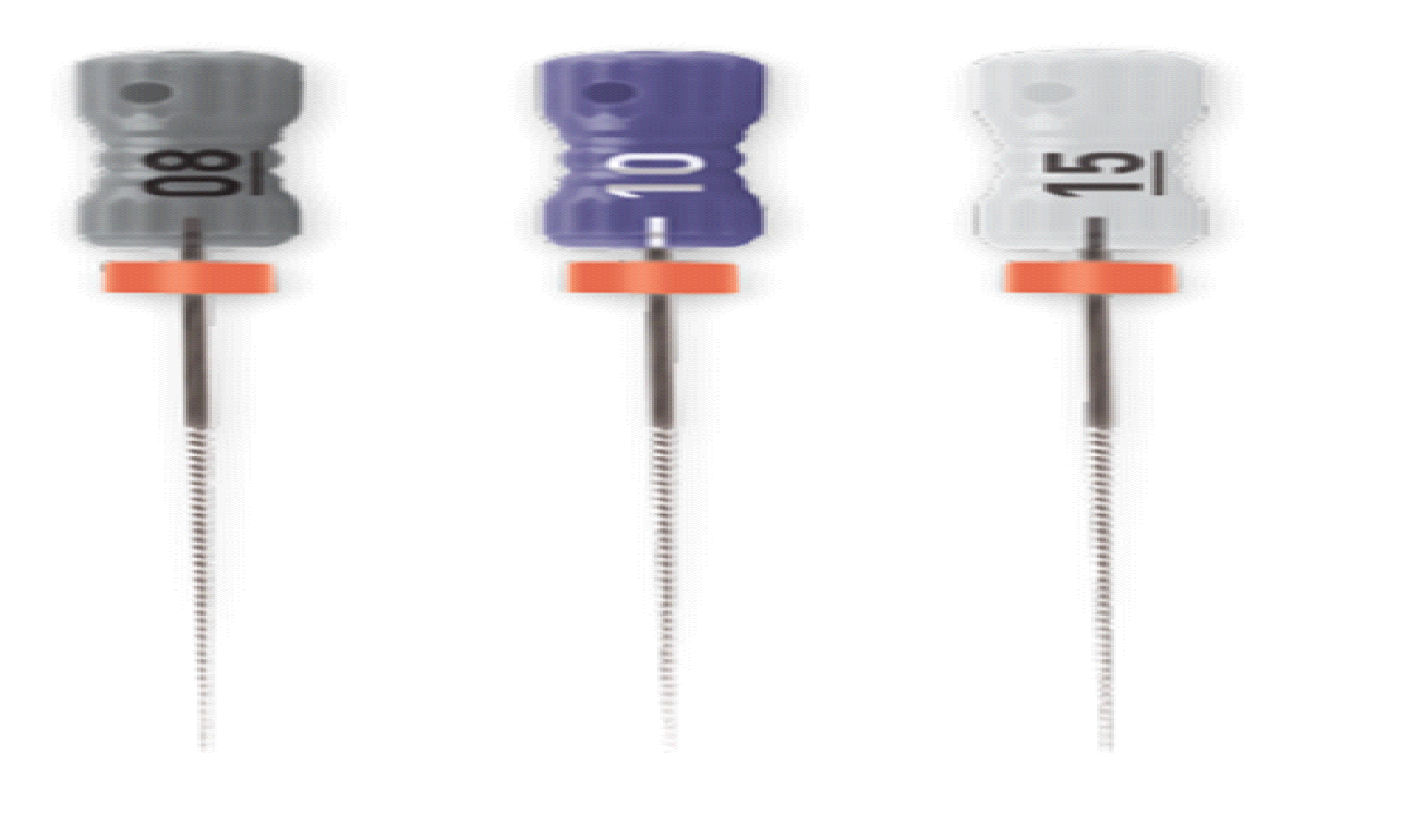
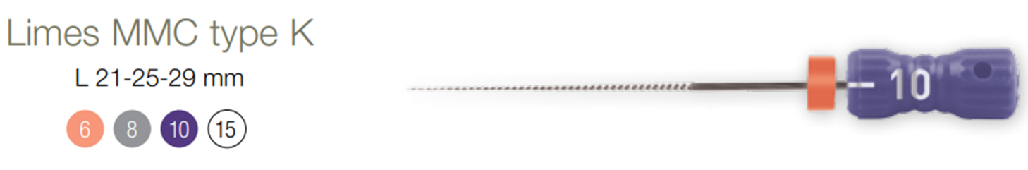
- MMC (micro-mega catheter)
- Obtained by cutting a cylindrical matrix, – Its section: hexagonal, – Intermediate instrument between a K file and an endodontic probe,
- A compact instrument, it only exists in the finest numbers: 06, 08, 10, and 15,
- Allows the first exploration of the channel
- Provides information on canal anatomy. The extent of mineralization of the canal lumen and the degree of curvature determine the difficulty of treatment and therefore the choice of the sequence best suited to the clinical case.
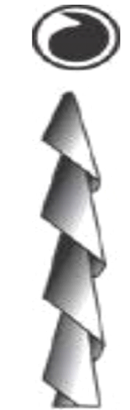
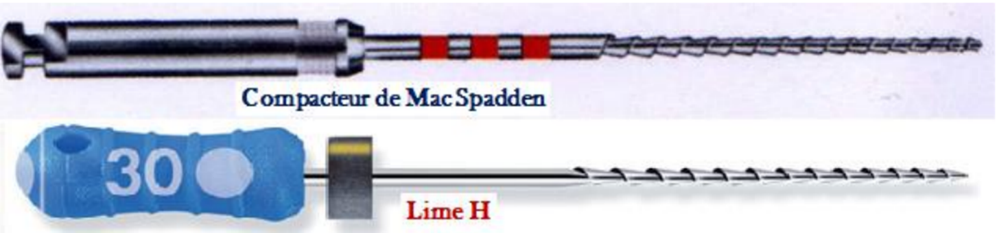
- Scrapers or File H (Hedström)
- Very sharp instrument, made from a round blank in the shape of an inverted Chinese hat,
- Identification symbol is a circle,
- Their sharpness is linked to their high angle of attack (90°). Their helix angle is 60°.
- Their step is short and constant,
- Their role is to broaden the preparation, while bringing up mineral and organic debris.
- Very fragile instrument, used only for traction; any rotational movement should be avoided.
- MME (micro-mega expander):
- It resembles the H file, but the angle of attack and the amplitude of its blades are moderate,
- It is an expanding instrument of No.: 08,10,15,
- A traction movement in the coronal direction,
- We use the MMC then the MME from No. 8 to No. 15.

Instrumentation in Endodontics
c. Reamers
- It is a standardized instrument that can be manufactured by twisting a triangular section blank or currently often machined directly.
- The identification symbol is a triangle.
- It has a long spiral pitch, therefore a low number of spirals (0.5 to 1 spiral/mm) with an average helix angle of 20°.
- The pin will be used by rotating it a quarter turn clockwise and then withdrawing it.
4.4. Ni-Ti hand instruments
– Have the advantage of being 6 to 8 times more flexible than traditional steel instruments
– These instruments manage to respect the anatomy and follow the initial canal path without the need for prior pre-curvature.
– However, Ni-Ti hand instruments have the disadvantage of lower cutting efficiency than those made of stainless steel.
– Today’s Ni-Ti hand instruments often have an active blade that is exactly the same as that of instruments designed for continuous rotation. Only their handle changes for manual gripping.
– They do not comply with the ISO standard and have different conicities greater than 2%.
Instrumentation in Endodontics
1.5. NiTi rotary instruments
a. Metallurgical characteristics:
– Super elasticity
– Flexibility
– Cutting efficiency
– Superior corrosion resistance compared to conventional instruments.
– Resistance to sterilization: sterilization does not affect the properties of NiTi.
- Hand-held NiTi instruments perform less well than steel instruments. Studies have shown that using these instruments in continuous rotation results in significantly higher cutting efficiency.
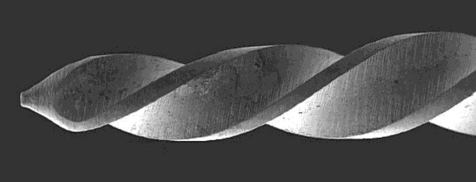
- Morphological characteristics:
- The active part:
- It differs depending on whether it is a passive instrument or an active instrument.
- For so-called passive or non-cutting instruments, the active part is fitted with a radial flat.
- For so-called active or cutting instruments: the active part does not have a radial flat

Passive or non-cutting instruments Active or cutting instruments
- The tip : it is blunt and not active and will serve as a guide for the progression of the instrument.
- The section : more complex than that of steel instruments.
- The angle of attack : it is either: – zero – positive – very positive.
- Taper : 4% or 6% can reach 12%
- Advantages of NiTi instruments: These instruments have enabled:
- An improvement in the quality of preparations
- Easy approach to complex cases,
- Improved coronal evacuation and less extrusion at the apical level help to minimize per or post-op risk
- Ergonomic and less tiring preparation due to:
- Well-established sequences
- On the mechanization of technology
- From the speed of the gesture.
- Limitations of use of NiTi instruments
- NiTi allergy;
- Oral access limited to these techniques, some manufacturers have developed specific rotating instruments (contra angle + instr) which are not bulky to remedy this problem (InGet system);
- Strong canal curvatures (risk of fractures which is controversial)
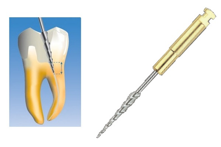
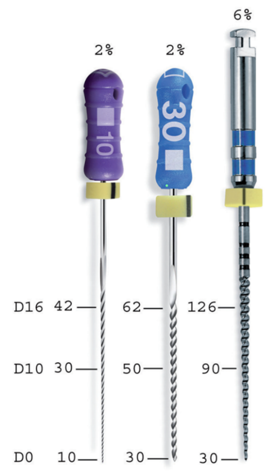
- Nickel-titanium instruments for opening the canal trajectory, or pre-enlargement
- In the canal shaping stage, two clinical problems arise recurrently:
– early blockage of fine and curved canals by creation of plugs or stops during catheterization with the successive back-and-forth use of manual steel files 08, 10 and 15;
– fracture of the tip of rotating nickel-titanium instruments.
– To overcome these problems, some manufacturers offer, in addition to the shaping sequences, rotating nickel-titanium instruments intended for early widening of the canal.
– These NiTi files, with 2% taper (Pathfile®, Dentsply Maillefer; Scout-RaCe®, FKG) or reverse taper (S-ApeX®, FKG), combine the rotational movement (which allows debris to rise and prevents blockages) with the flexibility of nickel-titanium in low taper (which prevents stops).
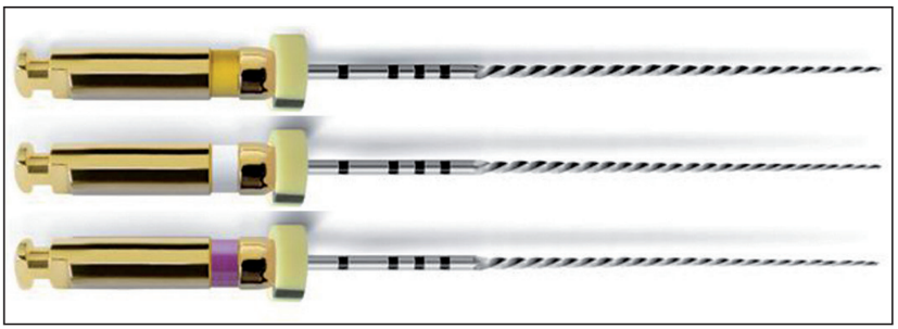
Instrumentation in Endodontics
PathFiles® pre-enlargement instruments, diameters 13 (purple), 16 (white) and 19 (yellow) in 2% taper. These instruments allow, after passing a small diameter hand file (08 or 10), to quickly open and secure the canal trajectory without risk of abutment or apical plug.
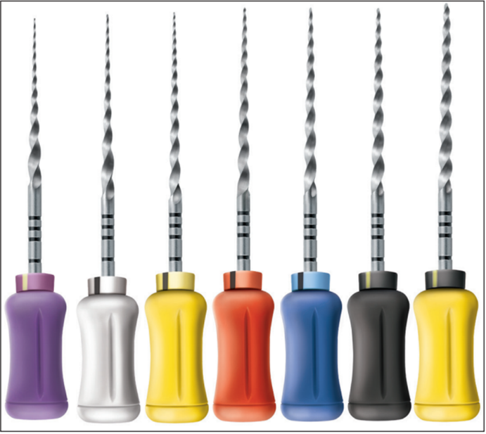
- Various mechanized systems
There are many mechanized Ni-Ti root canal preparation systems. Some classifications exist for them. However, none can ideally include all the techniques proposed as they are so numerous, varied and constantly evolving over the last fifteen years. These systems can be described by their indications, their manufacturing processes, their geometric characteristics and finally by the surface treatments they can undergo.
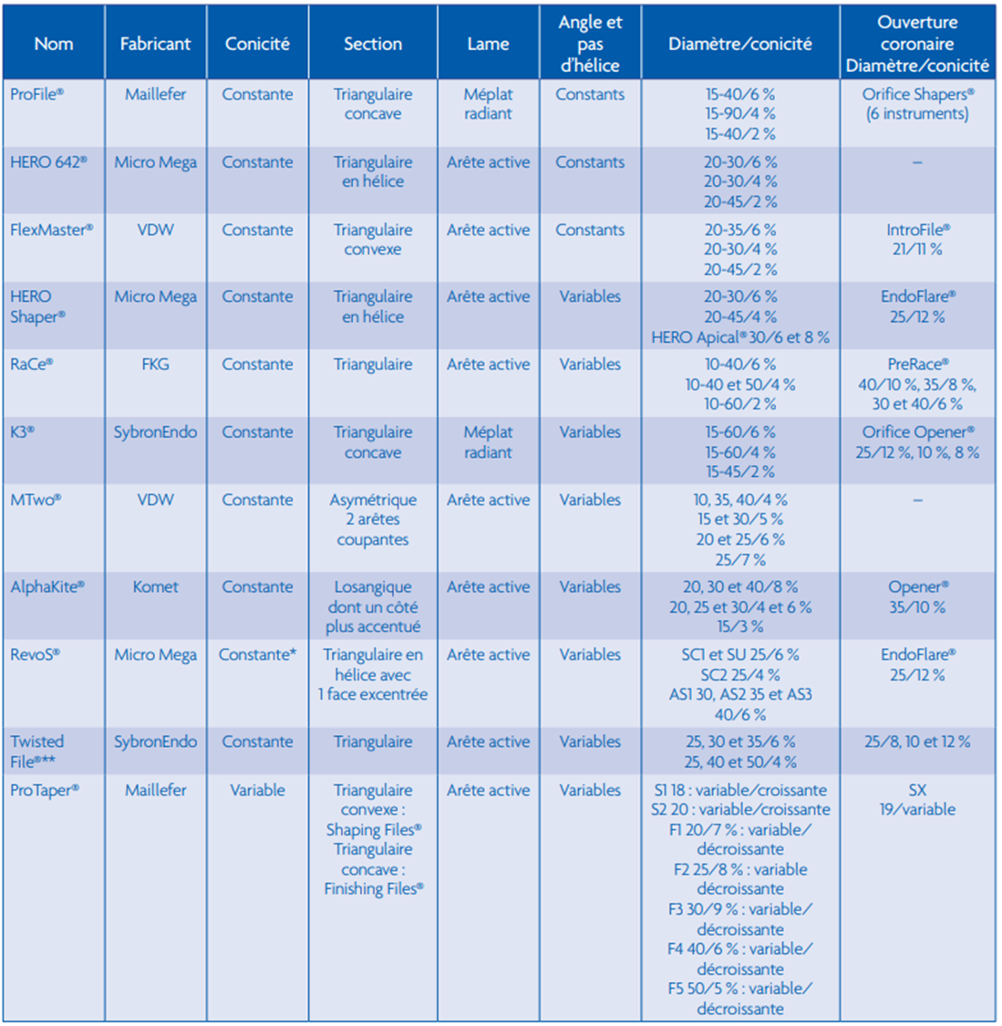
Non-exhaustive list of nickel-titanium instruments present on the French market and main characteristics
AS1, AS2 and AS3 have a 6% taper on the apical 5 mm then 0% taper on the rest of the blade.
** Twisted Files® are the only twisted (not machined) nickel-titanium instruments
Nickel-titanium instruments provide reliable and reproducible results if the following basic principles are scrupulously followed:
– the rotation speed recommended by the manufacturer must be respected by using contra-angles or specific motors;
– a nickel-titanium rotary file should never be inserted directly into a canal unless its patency has been checked with a steel hand file and it has not been pre-widened;
-the pressure on the contra-angle must be low and accompanied by a back and forth movement in the vertical direction, which allows the instrument to advance into the canal;
– after a few seconds of work, the instruments should be removed and wiped to avoid clogging of the coils, and the canal should be irrigated to remove suspended debris;
-instruments should never be kept rotating at the same length in the canal without vertical back-and-forth movement. Immobility in the vertical direction can lead to fracture of the instrument by cyclic fatigue or displacement of the canal trajectory with the appearance of a stop
-the instruments must be checked after each pass in order to detect any possible defect, precursor to fracture by torsion
- Material
The appearance of nickel-titanium instruments required the marketing and development of suitable equipment allowing their use at the appropriate speed.
There are currently several types of dynamic materials suitable for the use of nickel-titanium rotary instruments.
1 – Reducing contra-angles mounted directly on the chair
- Contra-angles reserved for nickel-titanium instruments have significant reduction factors (from 1/75 to 1/128). Some of them only offer a reduction while others combine it with a torque control function with automatic clutch, causing the instrument to stop if the pressure exerted during work is too great (NiTi Control®, Anthogyr; SiroNiTi®, Sirona).
- The clutch is supposed to reduce the risk of torsional fracture.
Most of these contra-angles have smaller heads than a classic blue contra-angle.
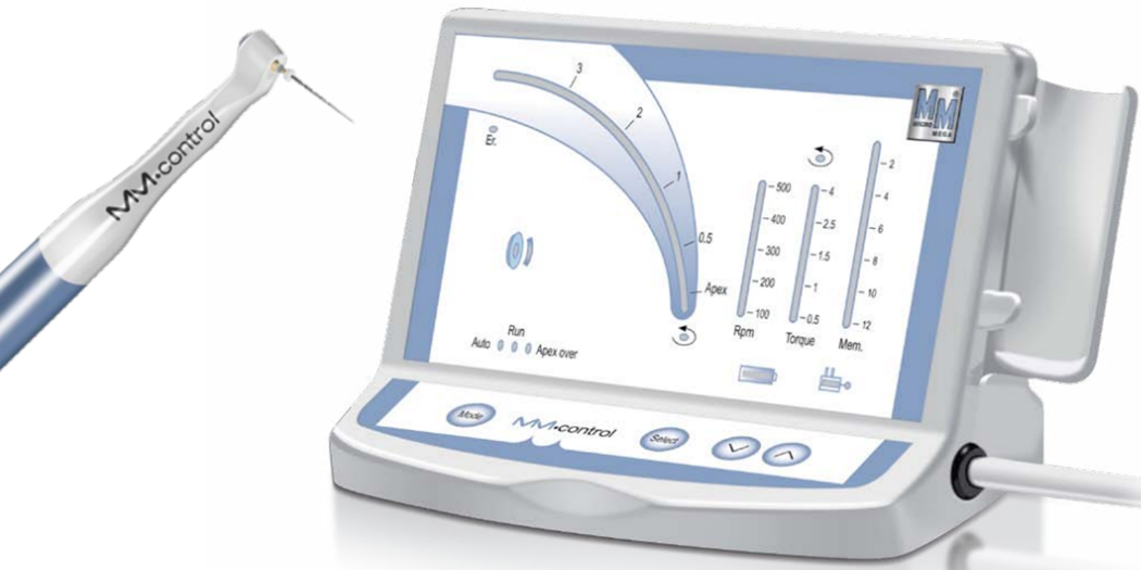
2 – Motors
The latest motors are compact, run on rechargeable batteries. They have torque adjustment options with a clutch and automatic reverse function (auto-reverse). They offer a double advantage: precise speed, electronically controlled and no vibration, noise or heating. However, they are an additional peripheral.
3 – Cordless motor contra-angle
- Some of these motors come in the form of contra-angles with a miniaturized motor housed in the handle. They have all the speed and torque adjustment features with automatic reverse function. They operate on rechargeable batteries and offer the undeniable advantage of being cordless, even if the maximum authorized torque is sometimes a little low. They nevertheless represent the solution of choice for warming up.

4 – Motors coupled to an apex locator
- Some manufacturers offer the combination of an endodontic motor with an integrated electronic apex locator and automatic reversal function.
- When the working length is detected by the apex locator, the contra-angle automatically disengages and begins a counterclockwise rotation. While these systems are ergonomically interesting at first glance, it must be kept in mind that an apex locator is never 100% reliable.
- Reciprocity
- Consists of animating a rotating NiTi instrument with a reciprocal movement, i.e. alternating the counterclockwise and clockwise directions of rotation with different rotation angles in order to eliminate the risk of screwing the instrument into the canal caused by the continuous rotation.
- The instrument first cuts counterclockwise and then disengages clockwise
- Counterclockwise rotation is greater than clockwise rotation: the instrument advances towards the apex
- The Wave One System
- The Reciproc system
The Wave One System
The Reciproc system
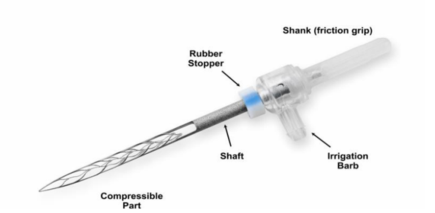
- Self Adjusting File (SAF) System = the self-adjusting file: a hollow, compressible, flexible, and deformable file
– The file adapts itself to the shape of the canal
– It is coupled with an irrigation system.
Instrumentation in Endodontics
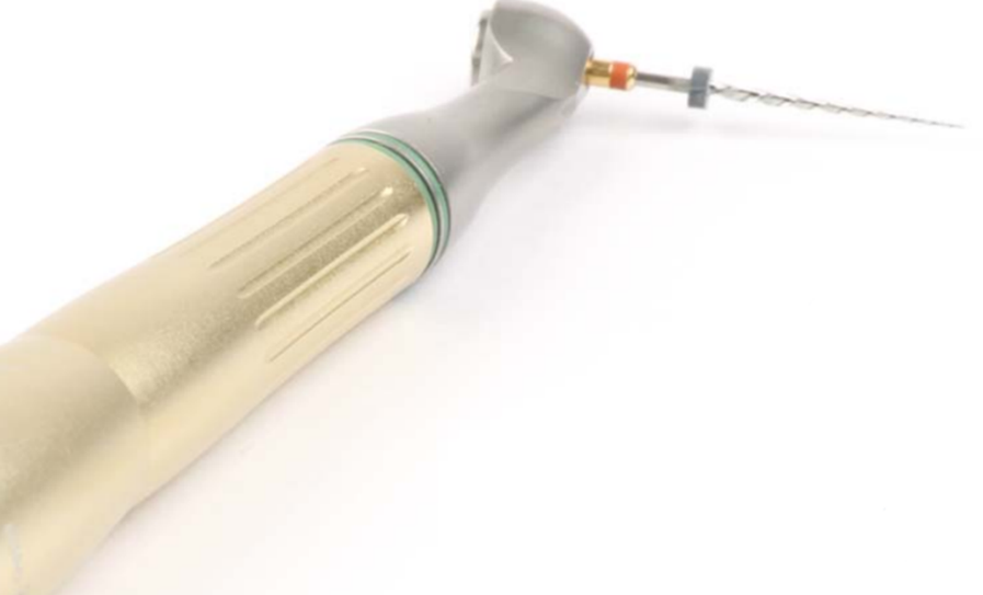
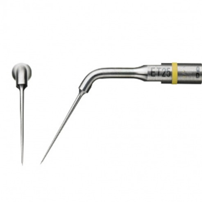
4.6. Sonic and ultrasonic instruments
– Various instrumentations are offered by the manufacturers.
– These are usually endodontic inserts mounted on ultrasonic generators.
– These systems constitute the best means of irrigation and sanitation of the endodontic cavity, they allow activation of the irrigation for better chemomechanical preparation and optimal elimination of the dentinal smear.
– Broken instruments can be recovered using this technique.

5. Instruments for root canal obturation
5.1. Manual instruments for root canal obturation
- “Spreaders” manual rammers for lateral condensation
These are pointed-end channel rammers.
They are intended to condense cold gutta percha laterally.
- Manual pluggers for vertical condensation “Pluggers”
- These are flat-ended conical rods for vertically tamping hot gutta percha.
- They are available in 2 shapes: Long handle; Short handle (Finger Spreader, Finger Plugger)
- Hand instruments for heating gutta-percha:

The “Heat Carriers”:
- They are pointed instruments that resemble a Rhein probe type probe.
- They are heated to red and carried into the canal to soften the gutta (which will be secondarily condensed with the pluggers).

5.2. Mechanized instruments for root canal obturation
- Rotating Pastry Cutters
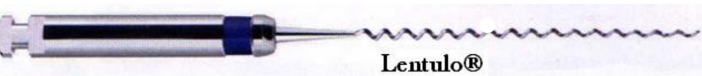
- They are of the LENTULO® (Dentsply Maillefer) or PASTINJECT® (Micro-Mega) type.
- The LENTULO®:
- Obtained by twisting a metal wire resulting in a very regular, thin and flexible screw,
- Used clockwise on a contra angle with low speed,
- PASTINJECT®:
- Same profile as the lentulo, but whose outline is a twisted flat blade and no longer a wire
- Very effective instrument, requires an excellent apical stop otherwise frequent overshoots occur.
– Paste fillers are used to carry out intracanal placement:


- Obturation pastes (in old obturation techniques based on the principle of paste alone or paste associated with a single-cone of gutta-percha ),
- Calcium hydroxide pastes (in inter-session fillings of endodontic treatments, in the treatment of necrotic teeth – balm categories IV).
Thermomechanical gutta-percha compactors
- Mac Spadden Compactor:
- It is a standardized instrument, used on a contra-angle for the obturation of root canals by thermomechanical condensation of gutta percha,
- It is made of stainless steel and is shaped similarly to an inverted H file.
- Their use requires training and a certain mastery.
- It is limited to the straight portions of the canal
- Recently Mac Spadden has been offering NiTi compactors, allowing the thermomechanical sealing of curved channels.
CONCLUSION
The new endodontic instruments are all finely designed and to provide the best performance, they must be used strictly and integrated into a method. The practitioner must not use one or the other of these instruments indifferently at the risk of incurring systematic failure but choose them according to an operative problem in a logical sequence.
Cracked teeth can be healed with modern techniques.
Gum disease can be prevented with proper brushing.
Dental implants integrate with the bone for a long-lasting solution.
Yellowed teeth can be brightened with professional whitening.
Dental X-rays reveal problems that are invisible to the naked eye.
Sensitive teeth benefit from specific toothpastes.
A diet low in sugar protects against cavities.